Microstructure and mineral chemistry
Thin-section investigations were addressed to characterizing the petrography and microstructure of the Antrona ultramafic rocks. The main rock types are: serpentinized peridotite (both foliated and mylonitic serpentinites), clinopyroxene-rich layers (infolded within serpentinite), chloriteschists, coarse-grained peridotite, dunite, and amphibole-rich layer. In all rocks we observed a main penetrative foliation that we called S2 on the basis of the proposed microstructural evolution. S2 foliation thus constitutes a fundamental reference point in the structural reconstruction.
In serpentinites, S2 foliation is mainly defined by serpentine. In some foliated serpentinites, S2 is marked by millimetric boudins or lozenge-shaped aggregates of fine-grained cpx+amp±chl+mag interpreted as deriving by replacement of earlier clinopyroxene, since relict clinopyroxene porphyroclasts are still locally recognizable inside the mineral aggregate. Consequently, the lozenge-shaped aggregates (see Fig. 2b), although now oriented parallel to S2 structures, may represent relict pre-S2 structures. Mylonitic serpentinites are made of serpentine, magnetite, and carbonate. Serpentine crystals with SPO mark the mylonitic S2 foliation often deformed by small-scale D3 crenulation (see also Fig. 2e). Ca-carbonate occurs as millimetric porphyroblasts showing intracrystalline deformation as well as smaller grains distributed in the serpentine groundmass.
In the cpx-rich layers transposed within the serpentinite (see Fig. 2c), the S2 is marked by fine-grained neoblastic clinopyroxene, sometimes with polygonal or mosaic-texture.
In chloriteschists, S2 foliation is defined by chlorite and amphibole crystals and is superimposed by incipient S3 foliation.
In coarse-grained peridotite and in dunites, S2 foliation is not visible and the static growth of chl±amp±srp is the only microstructure likely related to D2 stage.
The amphibole-rich layers consist of fine-grained aggregates of amphibole (~80%) and minor plagioclase and sphene oriented parallel to the S2 foliation. Few sub-millimetric anhedral crystals of amphibole or chlorite (pseudomorphs?) were observed in the fine-grained groundmass.
Post-S2 structures are represented by coarse-grained acicular crystals of serpentine or amphibole, which define an incipient and discontinuous S3 foliation, superimposed to the main foliation S2, and by veins. Veins are widespread in serpentinite and chloriteschists and are commonly filled with serpentine or chlorite fibres. All vein types crosscut all other structures, and are then later than pervasive S2 and incipient S3 foliations.
Microstructures older than S2 were observed in most fresh rocks and were used to unravel the oldest metamorphic evolution. These structures include S1 and pre-S1 structures.
S1 structures are mainly represented by relict foliations infolded within the S2 foliation. In serpentinized peridotites, S1 foliation is marked by sub-millimetric layers of neoblastic olivine and clinopyroxene replacing former olivine (Fo87-88) and clinopyroxene porphyroblasts (Wo50-En47-Fs3), respectively, still occurring in less recrystallized samples. These layers correspond to the brownish ol+cpx-layers recognized in the field and infolded within the serpentine S2 foliation (see Fig. 2a). In clinopyroxene-rich layers (see Fig. 2c), clinopyroxene shows three different textures: relict porphyroclast containing exsolution lamellae often filled with opaque minerals, mm-sized diopside porphyroblasts, often arranged in fan-shaped aggregates showing wavy extinction, and fine-grained granoblastic-neoblastic diopside, showing polygonal or mosaic-texture. Porphyroblastic diopside is here interpreted as relict S1 whilst porphyroclastic exolved clinopyroxene could be a pre-S1 structure.
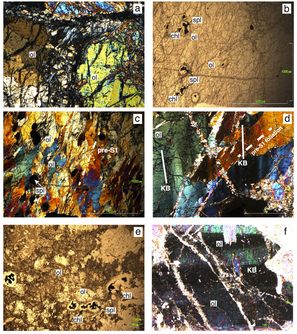
Pre-S1 structures are the oldest evidence of the tectono-metamorphic evolution recorded by the studied rocks and thus represent a key for unravelling the nature of their protolith (Fig. 3b and c). In serpentinites, pre-S1 structures are represented by: relict exolved clinopyroxene porphyroclasts (see above), olivine porphyroblasts, spinel porphyroclasts, and by lens-shaped or rectangular pseudomorphic sites of serpentine, probably replacing an earlier igneous mineral (pyroxene). In the least serpentinized samples, pre-S1 structure is attested by olivine occurring as round-shaped millimetric porphyroblasts (Fig. 3a). Olivine porphyroblasts commonly show intracrystalline deformation attested by the presence of wavy extinction and subgrain boundaries (or “kink-bands” = KB; Mercier and Nicolas 1975; Fig. 3a). Dunite layers and pods consist of olivine, spinel, opaque minerals, chlorite, ± serpentine (Fig. 3b, 3c). This rock is fine-grained, so no clear foliation is visible in the field. Under the microscope, dunite reveals the occurrence of a foliation marked by the shape-preferred orientation (SPO) of olivine porphyroblasts, commonly elongated and strained (Fig. 3c). Intracrystalline deformation is attested by the presence of kink-bands (“KB” in Fig. 3d). The angle between foliation and kink-bands boundaries (KBB) is about 45°. We interpret this foliation as a “pre-S1” structure. Olivine neoblasts in dunite grow at the rim of olivine porphyroblasts (both are Fo90), but more often they fill intercrystalline fractures in association with serpentine crystals (Fig. 3d). Spinel occurs as mm-sized holly-leaf shaped crystals or ovoidal porphyroclasts mantled by a chlorite rim (see Fig. 3b). BSE images of spinel (Fig. 4) show that in dunite spinel is characterized by a “porous” texture, as defined by Merlini et al. (2009). Spinel has a ferritchromite composition (Cr2O3 wt% ~ 55; FeOtot wt% ranging from ca. 35 to 37; see also Tartarotti et al., 2011). Element maps show the compositional variations of porous ferritchromite. The presence of Si, Mg, and scarce Al in the interstitial space visible in the maps suggest the occurrence of a silicate (serpentine? chlorite? undetectable due to its fine grain-size) forming intergrowths with spinel. Chlorite rimming the spinel crystals has Cr2O3 wt% content of up to 0.4; see also Tartarotti et al., 2011).
Coarse-grained ol+-cpx+-spl (±amp, ±chl)-rich rocks are characterized by olivine, clinopyroxene, pseudomorphic aggregates after probable orthopyroxene, and spinel (Fig. 3e). Olivine occurs as mm-sized olivine porphyroblasts (Fo87-88) showing intracrystalline deformation and olivine neoblasts (Fig. 3f), relict clinopyroxene porphyroclasts, and mm-scale chl+amp pseudomorphs (replacing orthopyroxene?). Spinel crystals are sub-millimetric and their shape recalls the holly-leaf habit described in mantle-derived peridotites (e.g., Mercier and Nicolas, 1975; Nicolas and Poirier, 1976). Spinel is always rimmed by a thick corona of chlorite. BSE images of spinel in these coarse-grained ol+-cpx+-spl-rich rocks (Fig. 5) show that spinel is characterized by a “porous” texture, similarly to the dunite spinel. Spinel has a ferritchromite composition (Cr2O3 wt% ranging from ca. 53 to 57; FeOtot wt% ranging from 35 to 39; see also Tartarotti et al., 2011). Element maps show the compositional variations of porous ferritchromite. The presence of Si, Al, Mg, Fe, and Mn in the interstitial space visible in the maps suggest the occurrence of chlorite, forming fine-grained intergrowths with spinel. Chlorite at the spinel rim has a Cr-chlorite composition (Cr2O3 wt% up to 1; see Tartarotti et al., 2011).
Figure 3. Photomicrographs of representative samples of the Antrona ultramafic rocks.
a) Olivine porphyroblasts (ol) with intracrystalline deformation; crossed nicols. b) General view of dunite (sample ATN64) with olivine and spinel porphyroblasts rimmed by chlorite; plane-polarised light. d) Elongate, coarse-grained olivine porphyroblasts (ol) showing intracystalline deformation, and spinel in fresh dunite (sample ANT64). Olivine shape preferred orientation almost coincides with rock foliation pre-S1 (see text); crossed nicols. d) Detail of dunite ANT64: coarse-grained elongated olivine porphyroblasts (ol) cut by veins filled with fine-grained olivine neoblasts. Orientation of internal kink-bands in olivine porphyroblasts (KB) describes an angle of about 45° with the rock foliation (pre-S1 foliation); crossed nicols. e) General view of olivine and spinel in coarse-grained ol+-cpx+-spl (±amp, ±chl)-rich rock (sample ANT120). Spinel is rimmed by chlorite corona; plane-polarised light. f) Detail of sample ANT120 showing kink-bands (KB) due to intracrystalline deformation of olivine porphyroblasts; crossed nicols.
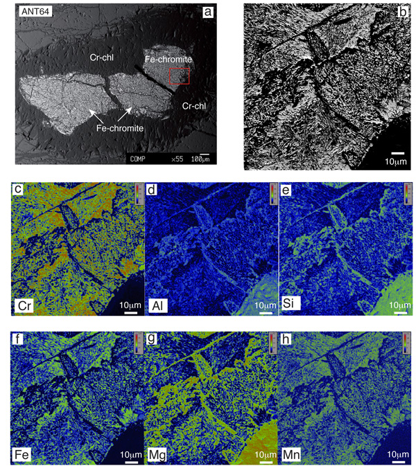
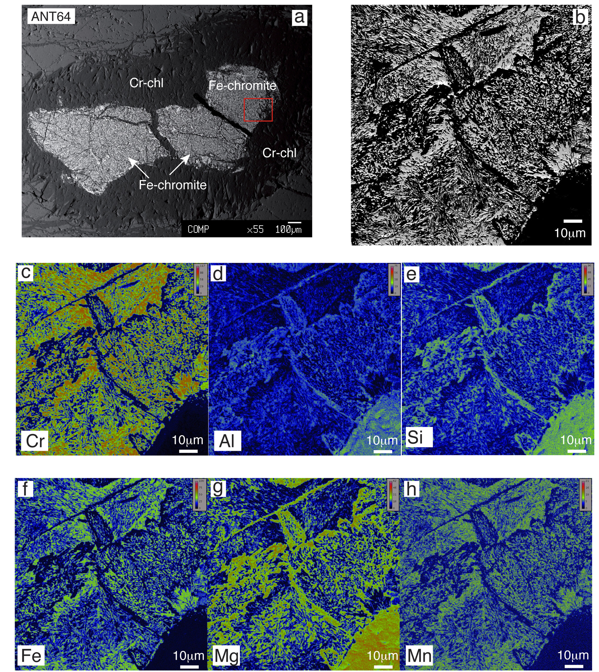
Figure 4. BSE images and X-ray element maps of spinel porphyroclast in dunite (sample ANT64).
Microchemical analyses of spinel are reported in Tartarotti et al. (2011). a) SEM image showing a general view of spinel (ferritchromite) porphyroclast rimmed by Cr-chlorite. b) Detail of picture (a) illustrating the “porous” texture of ferritchromite consisitng og fine-grained intergrowths of spinel and an unknown mineral. c-h) X-ray element maps showing the distribution of Cr, Al, Si, Fe, Mg, and Mn in the same crystal portion as illustrated in (b). Bright warm colors correspond to high concentrations; dark blue colors correspond to low concentrations. Ferritchromite is made of fine-grained intergrowths of spinel and an unknown mineral. The presence of Si, Mg, and scarce Al suggest that this mineral could be serpentine or chlorite; however it is not detectable due to its fine grain-size. Element maps performed at the by using a JEOL JXA-8200 probe, equipped with five WDS spectrometers and an EDS spectrometer, at the Dipartimento di Scienze della Terra “Ardito Desio” of the University of Milan.
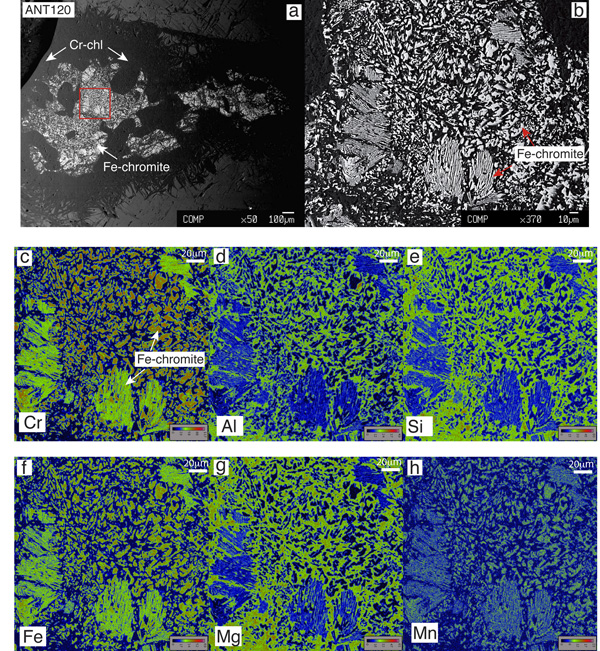
Figure 5. BSE images and X-ray element maps of spinel porphyroclast in coarse-grained ol+-cpx+-spl-rich rock (sample ANT120).
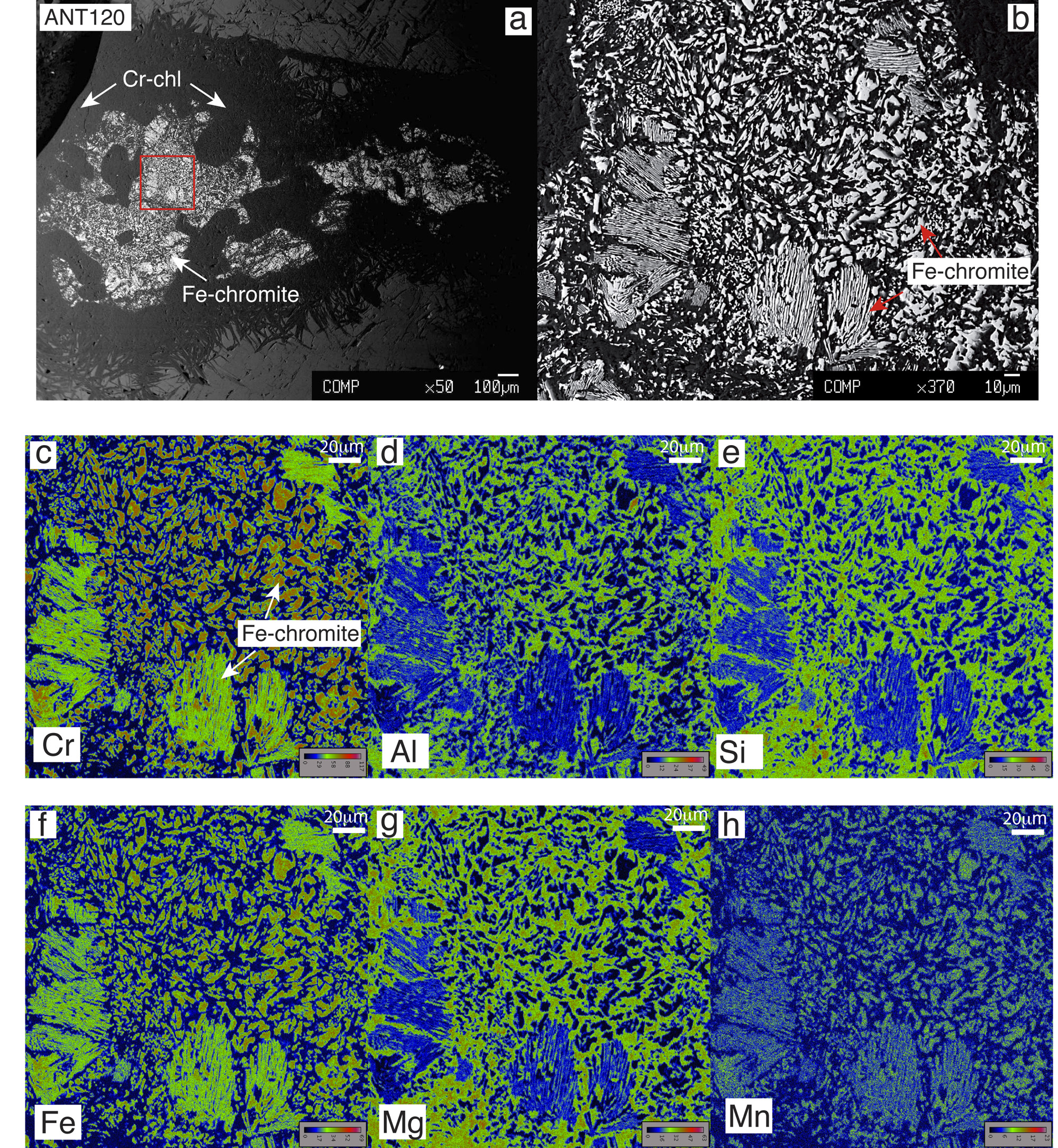
Microchemical analyses of spinel are reported in Tartarotti et al. (2011). a) SEM image showing a general view of spinel (ferritchromite) porphyroclast rimmed by Cr-chlorite. b) Detail of picture (a) illustrating the “porous” texture of ferritchromite consisting of fine-grained intergrowths of spinel and an unknown mineral. c-h) X-ray element maps show the distribution of Cr, Al, Si, Fe, Mg, and Mn in the same crystal portion as illustrated in (b). Bright warm colors correspond to high concentrations; dark blue colors correspond to low concentrations. Ferritchromite is made of fine-grained intergrowths of spinel and an unknown mineral. The presence of Si, Mg, and scarce Al suggest that this mineral could be serpentine or chlorite; however, it is not detectable due to its fine grain-size. Element maps performed at the by using a JEOL JXA-8200 probe, equipped with five WDS spectrometers and an EDS spectrometer, at the Dipartimento di Scienze della Terra “Ardito Desio” of the University of Milan.
The coarse-grained ol+-cpx+-spl-rich rocks are recrystallized at various extents into amp (tremolite)+chl-rich rocks, the resulting mineral assemblage and foliation thus depending on the extent of rock recrystallization.
Summing up, the porphyroblastic olivine, clinopyroxene, and spinel in less serpentinized peridotite, in dunite, and in coarse-grained ol+-cpx+-spl-rich rocks can be attributed to a pre-S1 structure representing the remnant of early, porphyroclastic mantle-derived texture (sensu Mercier and Nicolas, 1975). This pre-S1 structure has been overprinted by S1 structures as described above.