Itinerary 1: Nurra peninsula
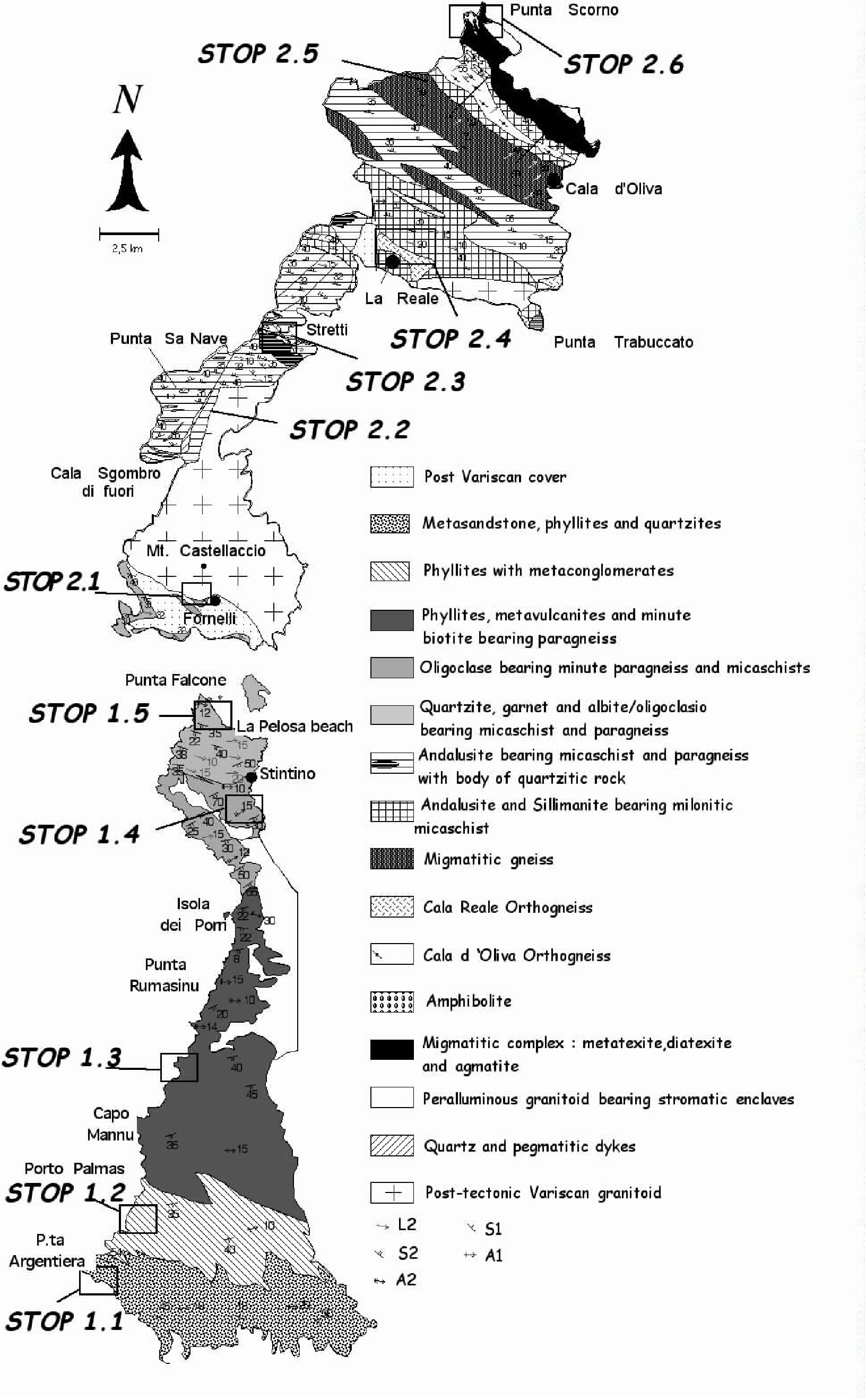
The first itinerary is dedicated to a cross-section through the Nurra peninsula starting from the southernmost and relatively less deformed area and going northward to Punta Falcone observing the increasing Variscan collisional deformation and Barrovian metamorphism in the basement (Figure 1.0).
Figure 1.0. Location of geological stops
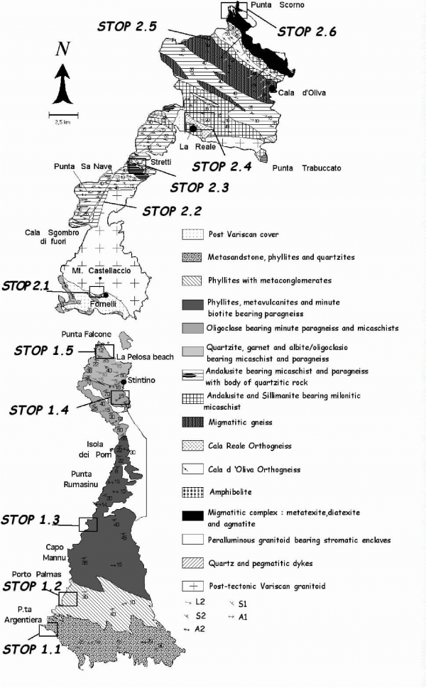
Location of geological stops of the itineraries in the Nurra peninsula (Itinerary 1) and Asinara Island (Itinerary 2).
Stop 1.1- Argentiera area - D1 Collisional deformation. (Coord. N040° 43’ 53.8’’; E008° 08’ 52.2’’).
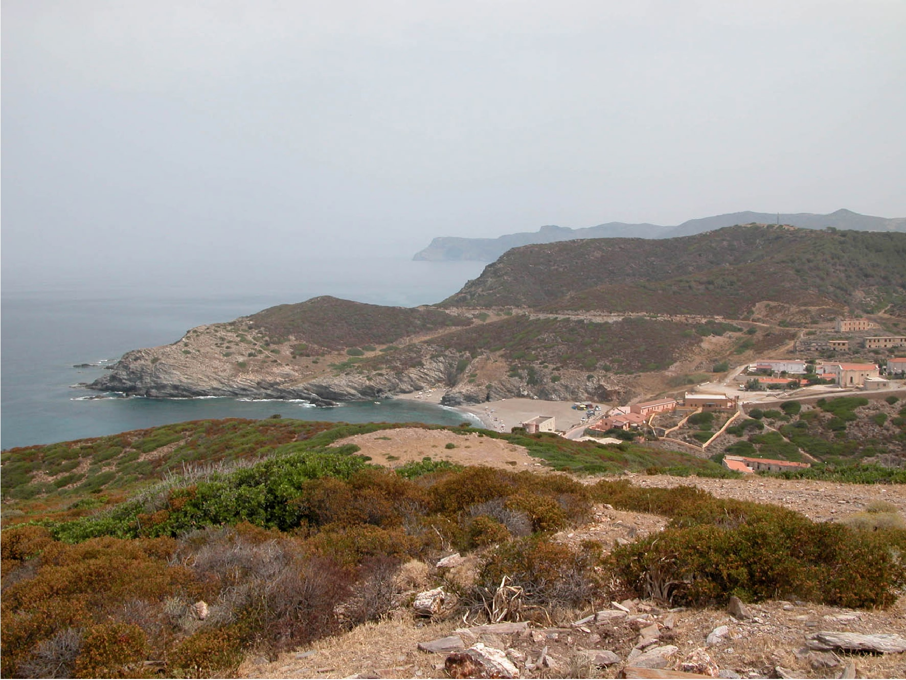
Figure 1.1. Overview of Argentiera mine
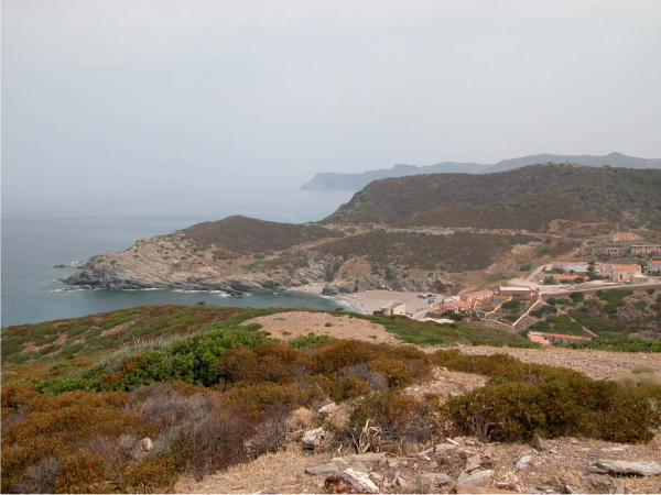
Overview of Argentiera mine and beach from the road going to Stop 1.1; view from south to north (Stop 1.1). In the far field low-metamorphic grade phyllites crop out along the western coast of the Nurra peninsula.
After reaching Palmadula small village we turn on the left toward Argentiera old mine and beach (Figure 1.1).
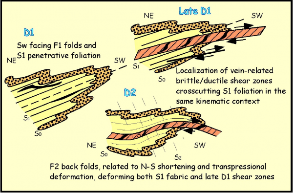
On the southern edge of Argentiera beach, the contact between pinkish ankerite bearing quartzites and blackish phyllites is exposed. In spite of the transposition of the primary contact, this stratigraphic boundary is well detectable and it is marked by a meta-diamictite of supposed glaciomarine environment of Hirnantian age (Oggiano & Mameli, 2006). This rock, when observed on sections parallel to the main foliation, appears as a dark laminated metapelite bearing sporadic angular clasts, millimetric up to metric in size, made up of quartzites and meta-sandstones. Moving towards the promontory of Punta Argentiera F1 recumbent folds, S1 penetrative foliation, S2 spaced crenulation cleavage, folds interference pattern and SW facing of F1 folds can be observed.
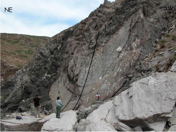
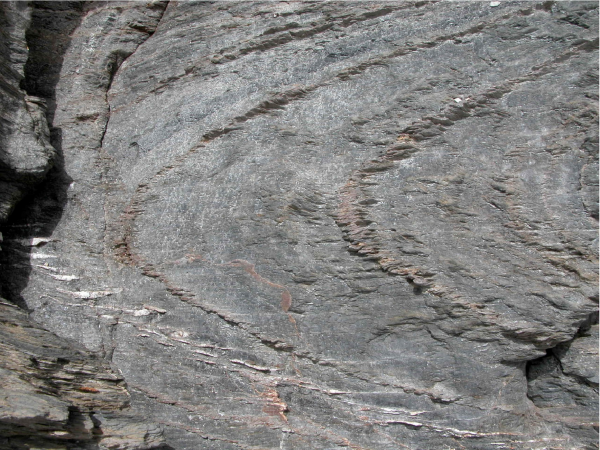
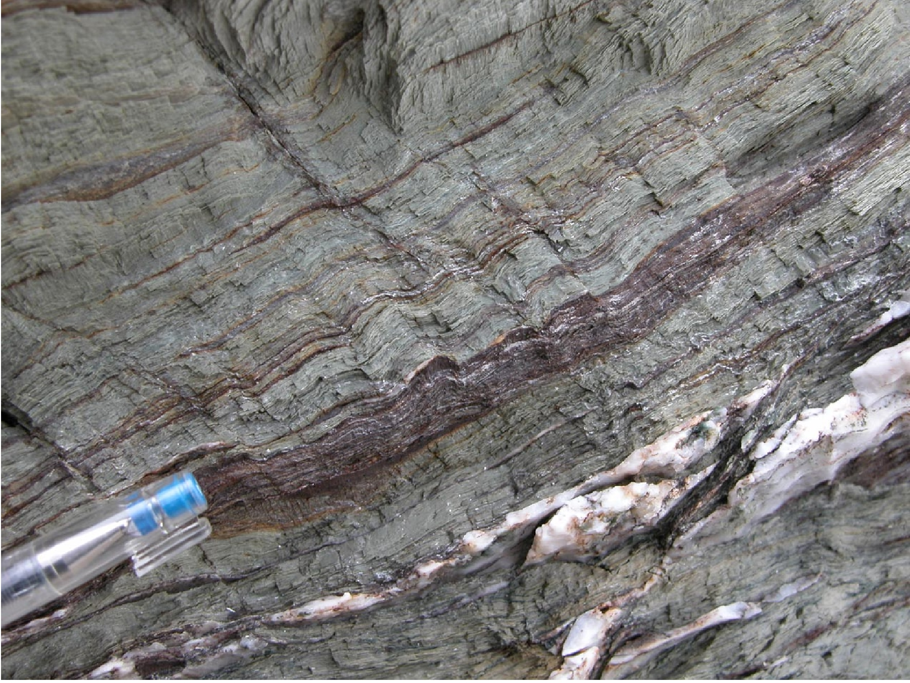
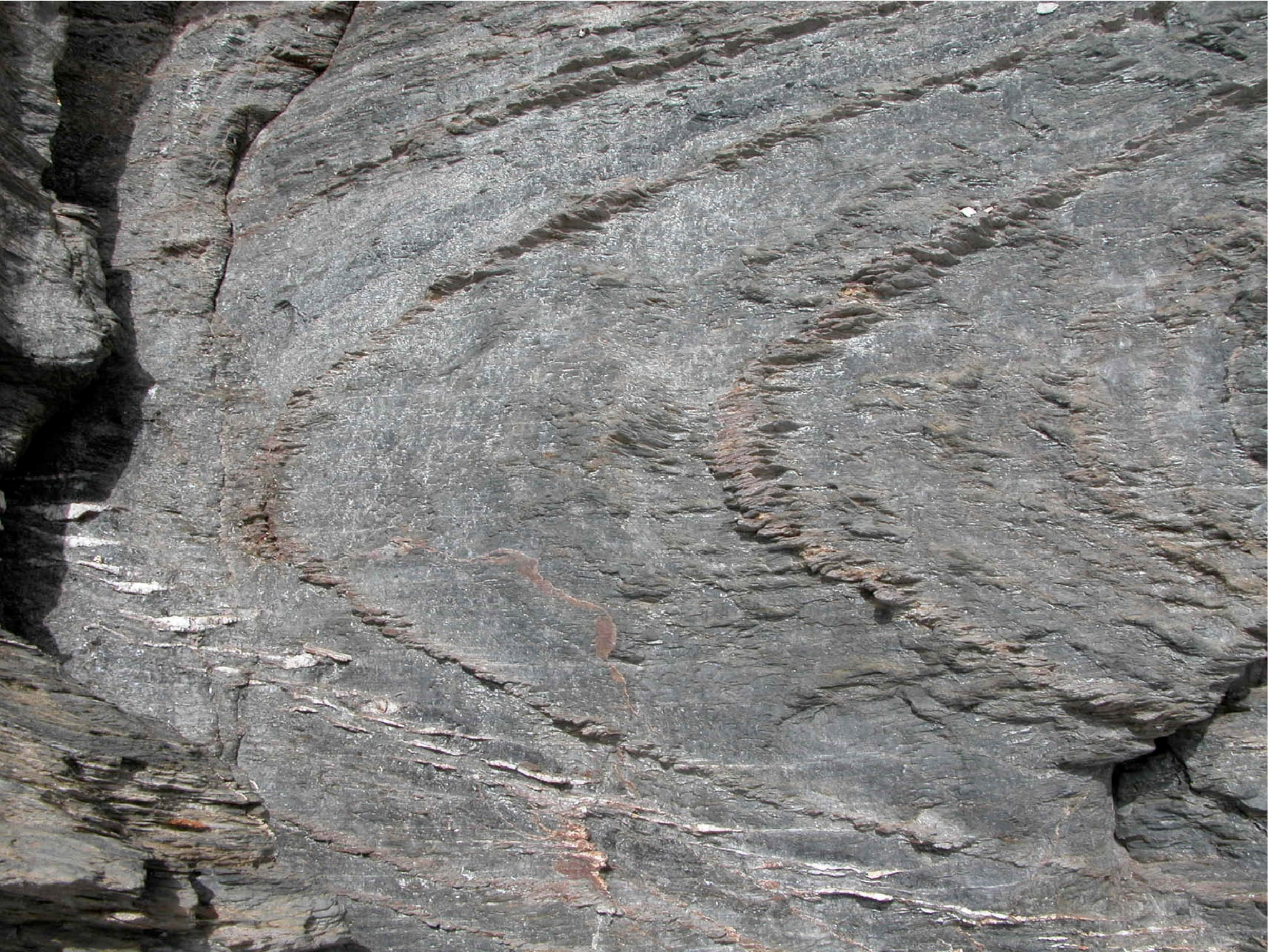
From the road going to the Argentiera beach we keep the road “via Pozzo Podestà”. We stop at a gate and after nerly 30 minutes of walking we descend a very steep slope for nearly 20 metres to reach the sea level at Punta Argentiera. Here, an hectometric- size F1 syncline affects phyllites and quartzites with nearly E-W fold axis (Figure 1.2). S1 axial plane foliation shows refraction through sedimentary layers. In very few places some load casts allow to detect the bedding polarity and the SW facing of the folds. The D1 structures are affected by open F2 folds and in places we can observe beautiful examples of interference patterns (Figure 1.3).

S2 is a spaced crenulation cleavage (Figure 1.4) dipping to the south. Locally we can see another crenulation cleavage conjugated to the S2 one. The outcrops are affected by several generations of veins, testifying a strong fluid activity during the deformation and prograde Barrovian metamorphism (Simpson, 1989) which here is confined within the chlorite zone of greenschist facies.

Stop 1.2- North of Porto Palmas. F1 folds, late D1 shear zones and veins; fluid inclusions informations, F2 open folds. (Coord. N040° 45’ 43.1’’; E008° 09’ 44.9’’).
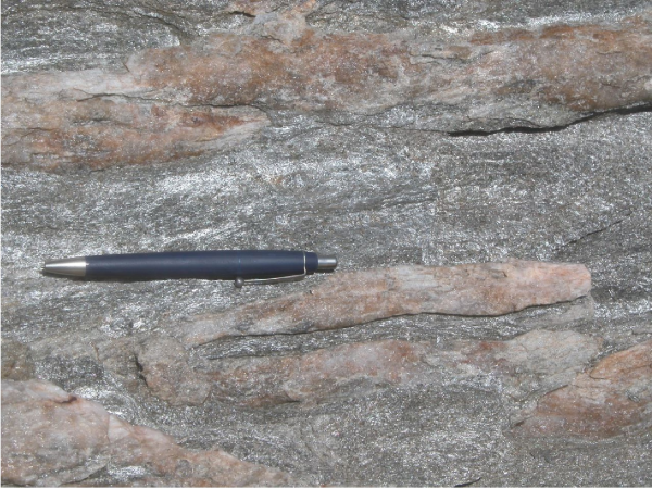
We come back from the same road until we reach the beach of Porto Palmas. Here we take a gravely road running northward for about 2 kilometers. At the sea level, below some meters of Quaternary deposits, several F1 tight folds crop out (Figure 1.5).
Figure 1.5. Mesoscopic tight F1 fold in metapelites
The folded surface is the sedimentary bedding and the sub-horizontal axial plane foliation is the S1 foliation. (Stop 1.2)
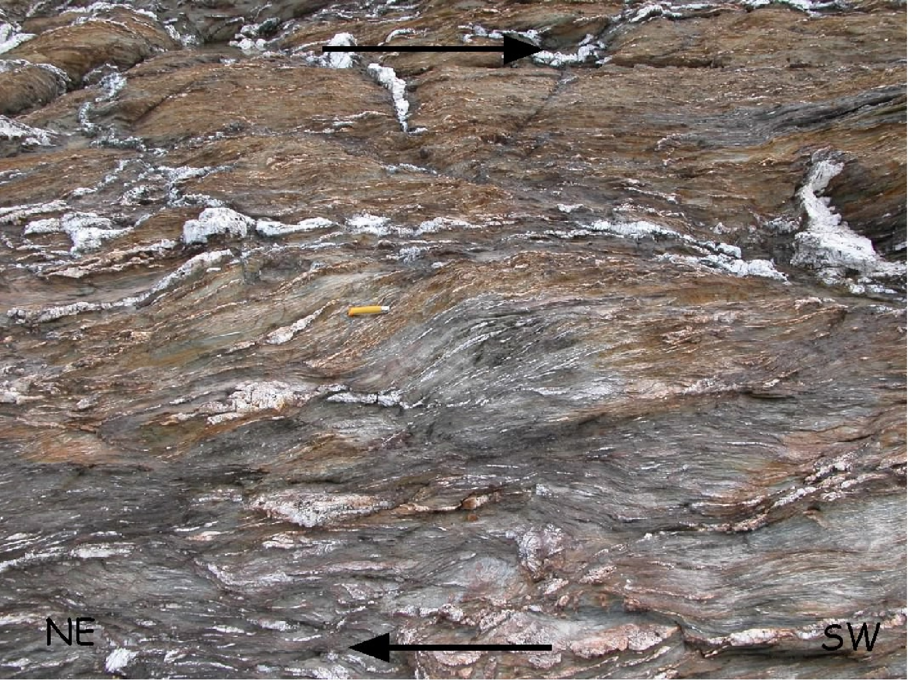
The sedimentary bedding is clearly visible at several localities highlighted by alternation of quartzites, metasandstones and dark phyllites and metapelites. Ductile-brittle shallowly dipping shear zones show a top-to-the S and SW sense of shear (Figure 1.6).

Overprinting criteria show that the shear zones deform the F1 folds that developed during the first D1 tectonic event, and are later deformed by the second D2 tectonic phase, associated, in the study area, with the development of a steeply dipping crenulation cleavage (S2).
Kinematic indicators, represented mainly by S-C fabric, point to a top-to-the SW sense of the shear, in accordance with the tectonic transport associated with the D1 event. These relations indicate that the development of shear zones may be confined to the final stages of the D1 tectonic phase, but before the D2 event (Figure 1.7).
Post-D1 flanking-structures, often of normal-type and associated to veins, are recognizable in the metapelites.
Microstructural analyses, on oriented samples of quartz veins from the shear zones provided informations on the relations between different fluid inclusions trapping events and the development of the shear zones. Sigmoidal quartz veins contains fluid inclusions trapped along secondary trails oriented both at high- and low-angles with respect to the shear zone boundaries. Both types of fluid inclusions contain an aqueous-carbonic fluids (Montomoli, 2003).
The ranges of the melting temperature of the carbonic phase (-56.5-57.4°C) and of clathrate (8.0-10.0°C) are similar in both inclusion types. On the other hand, the homogenisation temperatures of the carbonic phase are higher in type I (21.0-29.3 °C) than in type II inclusions (10.8-19.2°C). Raman analyses show that the two inclusion types usually have similar gas phase compositions (CO2=95.8-99.3 mole%, CH4=0.3-2.9 mole%, N2=0.2-1.3 mole%). Bulk compositions and densities have been calculated on the basis of the microthermometric and Raman data, and of the volume fraction of the gas phase. These calculations indicate that type I inclusions have lower densities (0.84-0.87 g/cm3) than the type II inclusions (0.88-0.92 g/cm3)(Figure 1.8). The computed isochores of both inclusion types do not match the P-T peak conditions proposed by Franceschelli et al., (1990) for the study area, suggesting that fluid inclusions have been trapped or re-equilibrated during a retrograde metamorphic pattern.
Figure 1.8. Fluid inclusions trapped along secondary trails
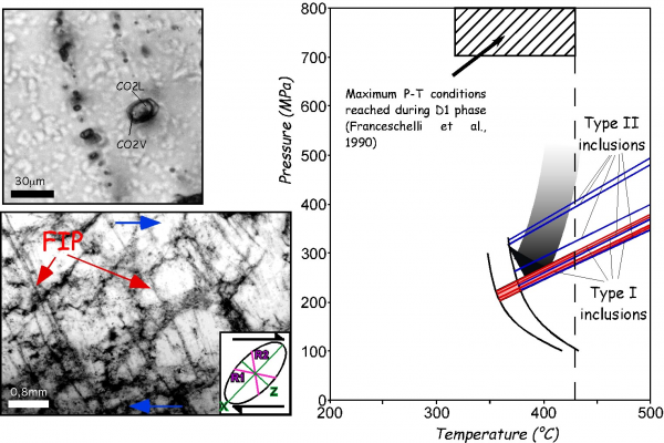
Fluid inclusions trapped along secondary trails related to non coaxial deformation in brittle ductile shear zones and possible P-T evolution defined with fluid inclusions isochors (Stop 1.2).
The geometry, kinematics and data on the thermo-baric evolution suggest that the shear zones developed in a compressive tectonic setting and played an important role in the exhumation of the low-grade metamorphic rocks of Northwestern Sardinia.
Stop 1.3- Villaggio Nurra: oolitic-iron deposits; metabasites.
From Porto Palmas we move to the north following the road from Palmadula to Stintino, then, turn to the west and after about two hundred meters, we stop at Villaggio Nurra, a bungalows turistic village. Here, on an overturned sequence, the contact between the Silurian black phyllites and the meta-greywakes, which host metabasites and metariolites, is exposed close to the cliff and in an old adit.
This contact is marked by an horizon of meta-diamictites made up of metapelites made up of chamosite in which fragments of quartzite and micaschist are scattered, revealing the recycling of a pre-Variscan basement. Other objects dispersed within the chamosite matrix consists of goethite pebbles, phosphorite nodules and chamosite ooids. Also this chemical sediment has been referred to the glacimarine setting which during late Ordovician extended over the north Gondwana shelf (Fadda et al., 2003).
The transition between upper Ordovician and Silurian is also characterised by the occurrence of alkaline to transitional metabasalts and metadolerites that occurs as sub-concordant sheeted dikes well exposed on the upper part of the cliff.
Stop 1.4- Road cuts in Le Saline area. (Coord. N040° 55’ 05.0’’; E 008° 13’ 48.8’’).
We go on to the north toward Stintino village. Nearly 2 km south of Stintino we stop at a road cut in Le Saline area. Micaschists and paragneisses are affected by close to tight F2 folds, testifying a northward increase of D2 deformation transposing the D1 fabric.
The metamorphic grade increases and we are near the zone of incoming of post-D1 garnets.
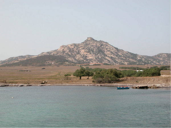
Stop 1.5- Punta Falcone (Coord. N040° 58’ 05.8’’; E008° 12’ 13.3’’).
We reach Capo del Falcone in the Roccaruja village area. Few meters above La Pelosa beach we can see northward in front of us the southern part of the Asinara Island. We can clearly recognize the granite of Mt. Castellaccio (pale yellow-pink) intruded in the metamorphites (lower dark rocks).
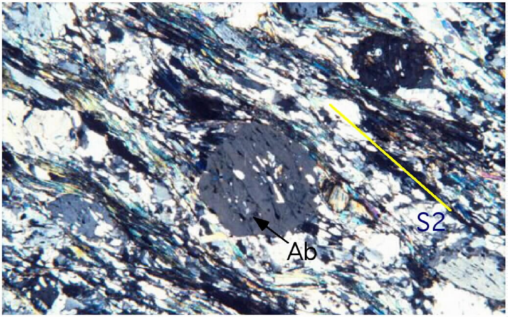
Albite and oligoclase bearing paragneisses and micaschists crop out. Plagioclase and garnet are of mm-size and visible with naked eyes. In thin section they preserve relics of S1 foliation and sometimes they are rotated during D2 with a top-to-NW sense of shear (Figure 1.9) and (Figure 1.10).
Figure 1.9. Syn-D2 partly rotated albite porphyroblast

Syn-D2 partly rotated albite porphyroblast (form Carosi & Oggiano, 2002) (Stop 1.5).
Figure 1.10. Rotated garnet (grt) porphyroblasts
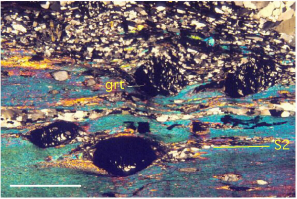
Rotated garnet (grt) porphyroblasts along the main S2 foliation, Scale bar is 2 mm (Stop 1.5).
The L2 stretching lineation is nearly parallel to F2 fold axes and strikes NW-SE. It is well-developed in quartz rods (Figure 1.11). Tight F2 folds (Figure 1.12) and sheath folds are recognizable in the paragneisses and in the transposed quartz veins. Beautiful examples of type-3 interference pattern between F1 and F2 folds are detectable in some road cuts.

Itinerary 2: Asinara Island
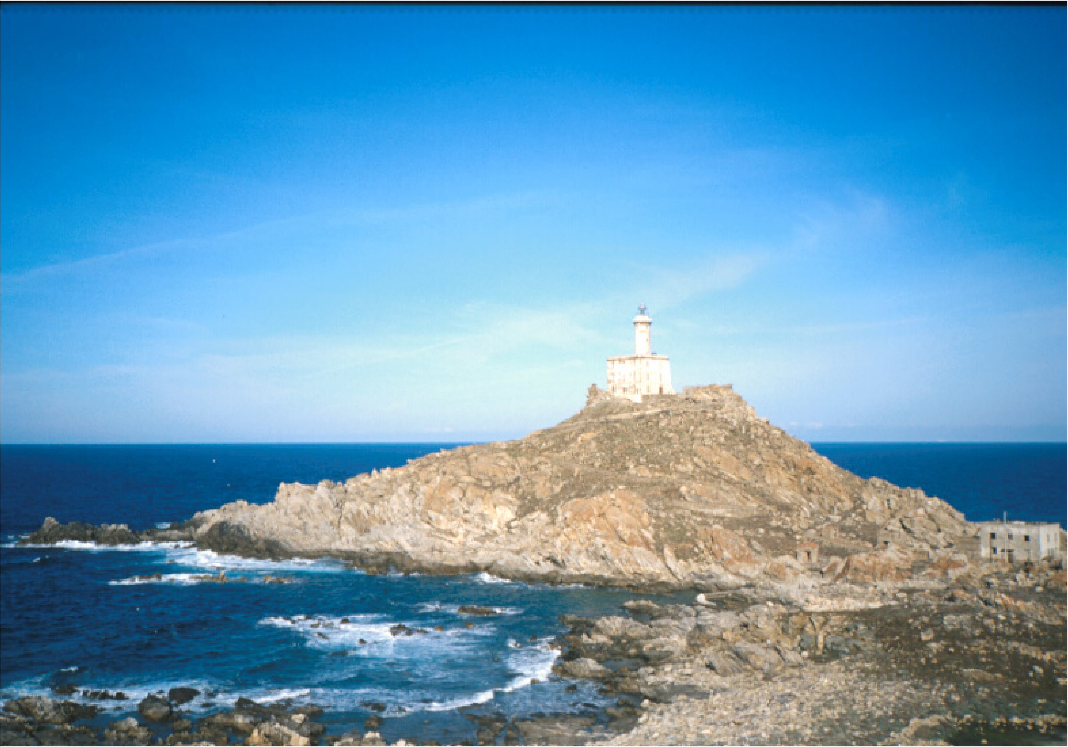
The second itinerary (modified after Carosi et al., 2004a, b) is entirely dedicated to the geology of the Asinara Island natural park. The itinerary starts at Fornelli, the point of berth of the ferry from Stintino (after 30-40 minutes of navigation), in the southernmost part of the island, and follows the main road till the old village of Cala d’Oliva to the north. From this old village the itinerary goes on by a badly preserved dirt road toward the Punta Scorno lighthouse, a beautiful wild place in the northernmost part of the island.
Asinara Island: historical background
Asinara is the second largest island of Sardinia after Sant\\'Antioco. It is a narrow territory stretching from North to South, with a very indented coastline, which highlights the great variety of habitats. The island has an extremely peculiar historical, environmental, and juridical situation.
We have only a few documents related to the last century, and they have been mainly filtered by the inaccessibility of the prison. What is sure is that the island has always been very busy for its barycentric position in the Mediterranean Sea. The Phoenicians knew it during their commercial routes, the Greeks used it as a landing port along their routes towards Provence, and also the Romans knew it during their sea adventures. The first Camaldolese monks reached it in the Middle Ages: they dedicated themselves to religious buildings and to the cultivation of the earth.
After the end of the great war, the island was divided among three Departments: the Navy Ministry for the lighthouses of Punta Scorno and the Reale; the Department of Health, from the Maritime Health Station of the Reale to Trabuccato; the Ministry of Justice, all the remaining territory being used as a working place in the open air. In the years after the Campaign of Ethiopia, between 1937 and 1939, hundreds of Ethiopian internees where deported to Asinara to be subject to “observation and health improvement”.
Although the first evidences of human presence date back to the Neolithic Age, the environmental features of the area have been preserved thanks to a series of events which gave it the name of "Isola del Diavolo" ("Devil\\'s Island"): it was a health center of quarantine, a camp of imprisonment during World War I, and one of the most important Italian superprisons during the terroristic period of the 70s and during the struggle against the organized crime, until the Park was established. This more than a century long isolation led on the one hand to the creation of the island\\'s aura of charm and mystery, and on the other hand to the indirect conservation of some integral and virgin areas which transformed it into an international unique and invaluable heritage.
Stop 2.1- Fornelli locality, nearby the southern contact between Mt. Castellaccio granitoid intrusion and the Medium Grade Metamorphic Complex. (Coord. N040° 59’ 37.9’’; E008° 14’ 09.4’’).
The Late Variscan Mt. Castellaccio intrusion consists of inequigranular granodiorite - monzogranite characterised by large laths of K-feldspar highlighting a magmatic fluidality. Small bodies of nearly equigranular and muscovite-rich monzogranitic facies also occur within the main intrusion (e.g. Cala S. Andrea) and in the northernmost part of the island.
Figure 2.1. Mt. Catellaccio
Mt. Catellaccio: the pinkish rocks is the granitoid whereas the dark rocks in the lower left part are porphyroblastic paragneisses (Stop 2.1).
Oligoclase bearing paragneisses and micaschists are mainly affected by the F2 tight to isoclinal folds and show the same structural and metamorphic history of the metamorphites cropping out at Punta Falcone.
It is worth to note that the effect of contact metamorphism on the intruded schists is very weak. Only in thin section recrystallization of biotite into decussate texture and destabilization of garnet is detectable. Several brittle shear zones and faults affect the metamorphites but for the lacking of post-Variscan sediments does not allow to attribute them to the Variscan or to the Alpine cycle.
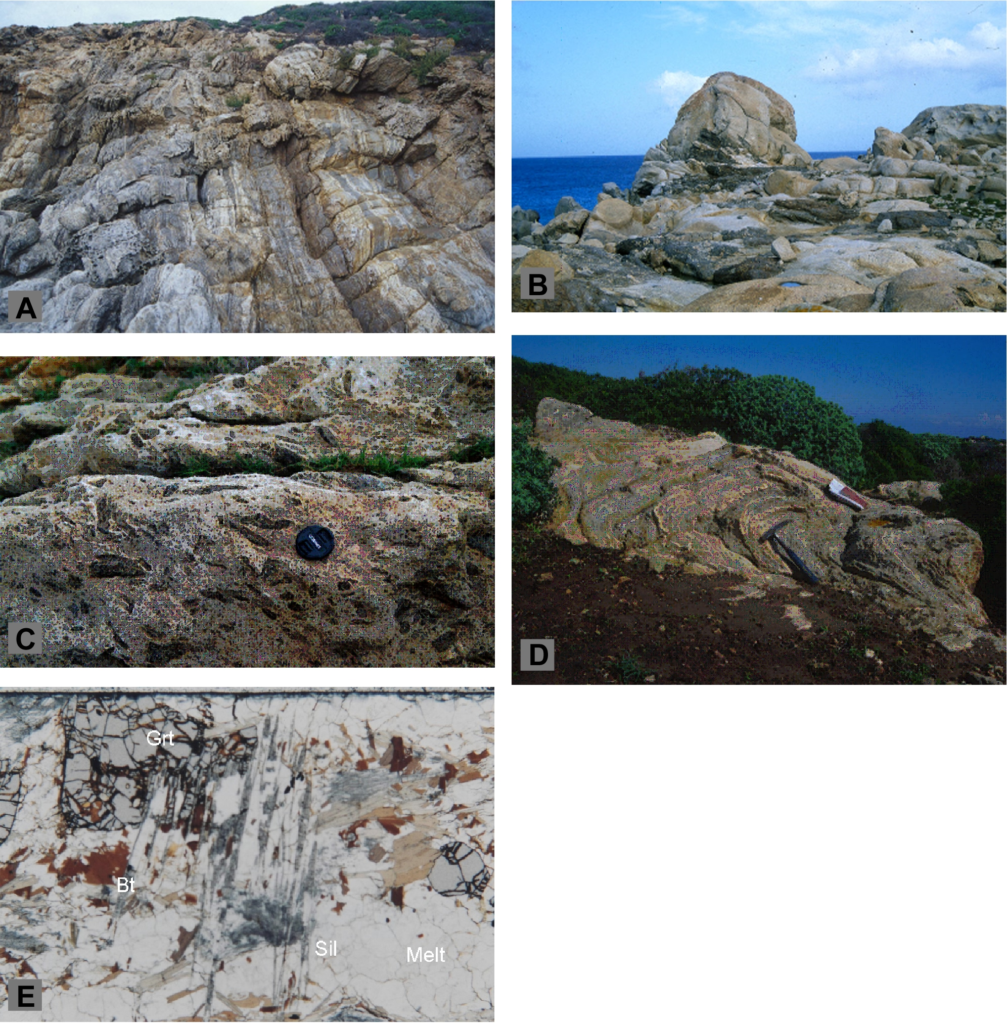
Stop 2.2- We go ahead to the north along the road to Cala d’Oliva and we stop few dozen meters after the northern contact between the granitoid intrusion and the Medium-Grade Metamorphic Complex (MGMC) in Punta Sa Nave and Stretti zone. (Coord. N041° 02’ 50.8’’; E008° 14’ 50.2’’).
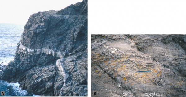
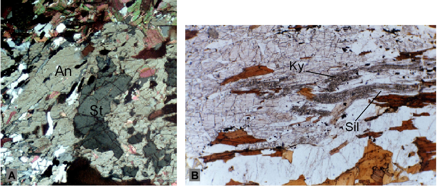
Here, in contrast to the southern contest, the MGMC schists are affected by an HT-LP metamorphism which overprints the previous Barrovian type one, giving rise to huge andalusite and sillimanite blastesis ± cordierite. We can observe andalusite-bearing paragneisses and micaschists; at places centimetric blasts of chiastolite are also present. This new assemblage overprints Grt + Sta (Kretz, 1983) and, at place also disthene, were stable (Figure 2.2A and B); relics of these minerals are still well detectable within the HT-LP related assemblages suggesting the following andalusite making reactions:
Figure 2.2. The HT-LP metamorphic overprinting (Stop 2.2)

A. Andalusite (An) overgrowth on the staurolite (St) relics in andalusite bearing micaschist (CPL, magnification 20x); B. Disthene (Ky)-sillimanite (Sil) showing subdivision of single kyanite blast into several sillimanite crystals (CPL, magnification 20x).
Sta + Mus = And + Bt + H2O (Figure 2.2A),
Sta + Grt + Qtz = Crd + H2O
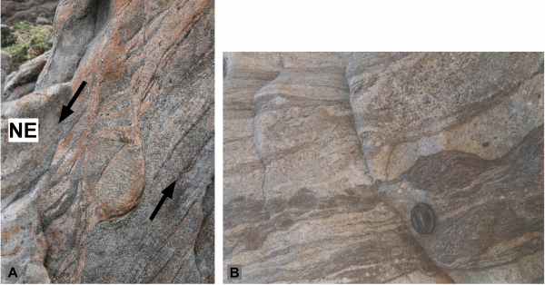
Moving some hundred metres northward, a horizon of massive quartzite crops out close to an alignment of hectometric to metric lenses and boudins of massive amphibolite. Relics gabbroic and basaltic texture joined to N-morb type signature according to Cappelli et al., (1992) led to consider these rocks as ophiolitic remnants. A Sm/Nd isochron on this amphibolite yields a late Proterozoic age. The two main deformation phases, D2 and D3, give rise to interference patterns visible both on the road cut and in the flat areas around. The main fabric is due to D2 deformation phase, but differently from the area south of the Mt. Castellaccio intrusion F2 folds verge toward the SW (Figure 2.3) and S2 schistosity dips northward.
Figure 2.3. Open to closed D2 folds

Open to closed D2 folds (sensu Fleuty, 1967) with a vergence to the south (Oligoclase bearing paragneiss, Stretti zone) (Stop 2.2).
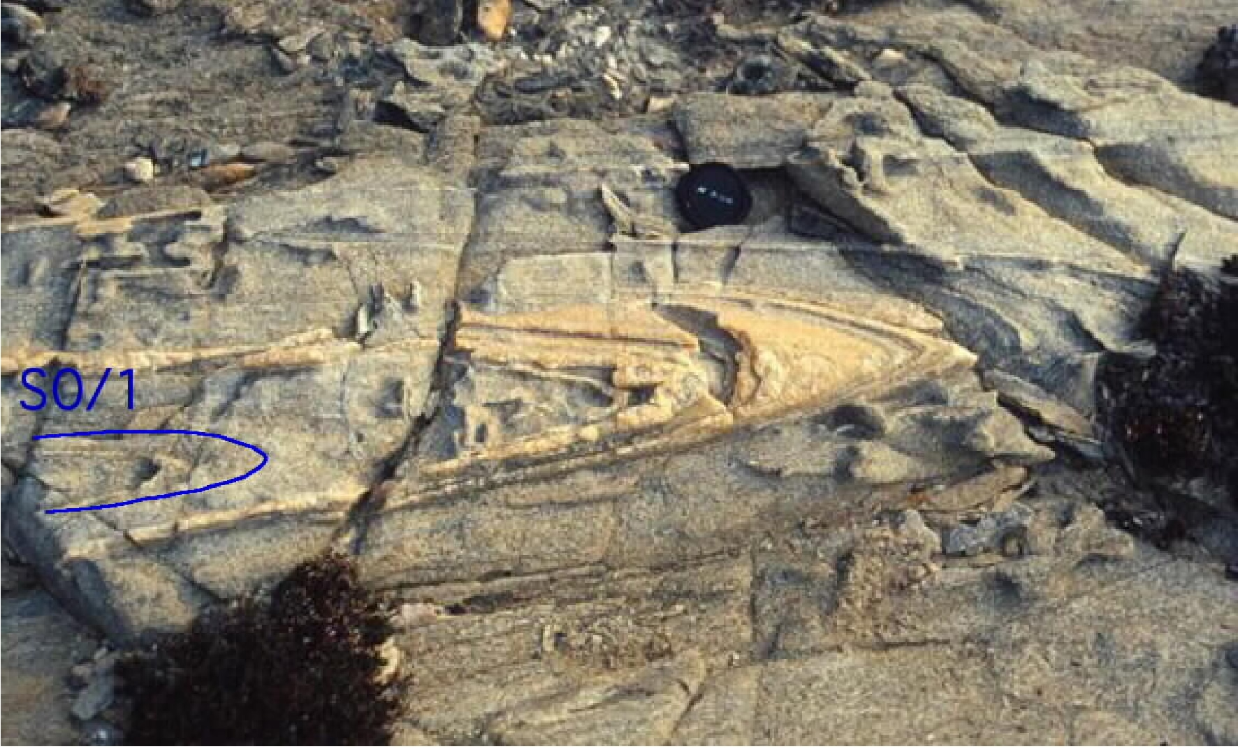
In the Stretti zone clear relationship between S1, parallel to S0, D2 tight folds and upright D3 folds can be observed. Pegmatite dykes intrude the MGMC both sub-parallel to the S2 schistosity and crosscutting the schistosity (Figure 2.4A) suggesting the existence of at least two intrusive events.
Figure 2.4. Stretti zone (Stop 2.2).
A. Pegmatitic dyke cross cutting at low angle the micaschist host rocks. Punta sa nave; B. Static large-size andalusite crystals in the micaschist (MGMC, Stretti zone). Pencil for scale.
In the Stretti zone the HT-LP overprint is characterized by static crystallization of cm-size andalusite crystals (Figure 2.4B) overprinting the D3 deformation phase. The L2 stretching lineation is well developed in the quartzites and it is nearly parallel to the F2 fold axes.
Stop 2.3- La Reale. (Coord. N041° 04’ 00.2’’; E008° 17’ 34.3’’).
Few dozen of meters behind the houses of La Reale, taking the road to the “diramazione campo faro” toward the so-called military cemetery, an orthogneiss body crops out. The prominent linear fabric is defined by laths of alkali feldspar in a matrix characterised by a preferred orientation of quartz, plagioclase, microcline, and muscovite weakly fibrolitized. The fabric is apparently referable to a L-tectonite. However, a closer look allows us to recognize that this is due to the interference between the main fabric (S2) and small-size collapse folds (with sub-horizontal axis and axial plane) related to a post-D3 nearly vertical shortening.
The High Grade Metamorphic Complex
Stop 2.4- From Cala d’Oliva to Punta Scorno. (Coord. N041° 07’ 03.3’’; E008° 19’ 04.0’’).
We continue northward until we reach Cala d’Oliva and we follow a gravely road on the left climbing a small hill and we descend toward Punta Scorno in the lighthouse area. We crosscut the High Grade Metamorphic Complex, mainly composed by migmatitic gneisses, orthogneisses, metatexites and diatexites.
We make some short stops along the road to observe the Cala d’Oliva orthogneiss and andalusite bearing mylonitic micaschists (Figure 2.5).
Figure 2.5. Example of andalusite and fibrolite bearing mylonitic micaschist

Example of andalusite and fibrolite bearing mylonitic micaschist (HGMC, Punta Soriana, central Asinara)(Stop 2.4).
The Cala d’Oliva orthogneiss is a sheet gneiss body with variable thickness defined by plagioclase-antiperthite and microcline-perthite interlayered with discontinuous domains depleted in feldspars and enriched in phillosilicates. During the HT-LP evolution such domains experienced widespread static blastesis defined by cordierite, sillimanite and andalusite. The new static assemblage partially erased a mylonitic fabric which is still documented by annealed quartz ribbons. On the prominent schistosity pronounced stretching lineation with a down-dip attitude is well exposed.
Going northward we enter in the migmatitic complex made up of metatexites (Figure 2.6A), diatexites characterised by either stromatic, nebulitic and agmatitic textures with sometime restitic biotite enriched domains (Figure 2.6B and C). In the neosome migmatite portions clear blastesis of static biotite and prismatic silliminate could be observed (Figure 2.6E). In the metatexitic complex nice examples of D4 structures (tension gashes, flanking folds) are recognizable (Figure 2.6D).
Figure 2.6. The Migmatitic Complex (Stop 2.4).
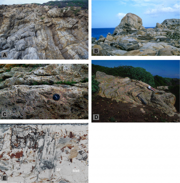
A. Strongly deformed metaxite with main foliation parallel to the leucosome-mesosome boundaries.; B. Anatectic intrusive complex enclosing bodies of melanosome and paleosome; C. Leucogranite body with relics of restitic paleosome and schlieren structures (Punta Sabina); D. Metatexite complex with late D4 fold showing a sub-horizontal axial plane (road to Punta Scorno); E. relic of atoll- like garnet (Grt), biotite (Bt) within leucosome melt (quartz+plagioclase) and late prismatic sillimanite (Sil) (Diatexite). PPL, Magnification 20x).
Close to Punta Scorno the HGMC is defined from south to north by mylonitic micaschists, banded amphibolites, orthogneiss and sheared metatexites and diatexites.
The Punta Scorno orthogneiss, normatively a syenogranite, consists of mylonitic augen gneiss characterised by large laths of alkali feldspar which exhibit both oblate and prolate shapes. The mineral assemblage is the same of Cala d\\'Oliva orthogneiss even if mica, garnet and sillimanite are comparatively more abundant. It is characterized by cm- to dm-size laths of k-feldspar with asymmetric recrystallized tails (visible at the naked eye) pointing out a top to the SW sense of shear with a down dip stretching lineation (Figure 2.7). Sometimes such fabric is overprinted by a late D3 crenulation (Figure 2.7). Nice examples of rotated porphyroblasts both in the XY and XZ principal planes of the strain ellipsoid with well developed quartz tails are observable in some outcrops at the sea level on the left side of the road.
Figure 2.7. The Punta Scorno orthogneiss (Stop 2.4).
A. Recrystallized tails on the S2 foliation. Orthogneiss, HGMC; B. D3 crenulation of shear sense indicators in the Punta Scorno orthogneiss (HGMC) observed on a plane parallel to the lineation and orthogonal to the foliation. Shear sense is top-to-the SW.
F3 nearly upright folds affect the amphibolites and the surrounding rocks. It is easy to observe refolds F2 folds and fold hinges.
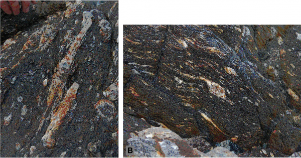
Hectometric-size outcrops of banded amphibolites are visible on the northwestern coast (Figure 2.8) south of the lighthouse and consist either of centimetric to decimetric dark amphibolites alternated with leptinitic layers, or massive hornblende-plagioclase amphibolites. The mineral assemblages sometime include calcic–pyroxene and garnet (both as relics of previous granulite metamorphic stage) and zoisite.
Figure 2.8. Banded amphibolite

Banded amphibolite embedded in the migmatite complex. Punta Scorno Area (Stop 2.4).
Stop. 2.5- Punta Scorno.
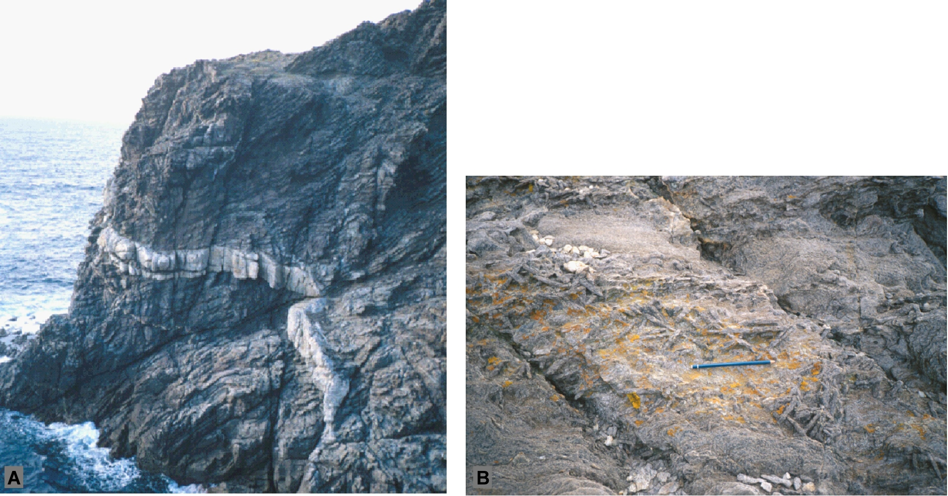
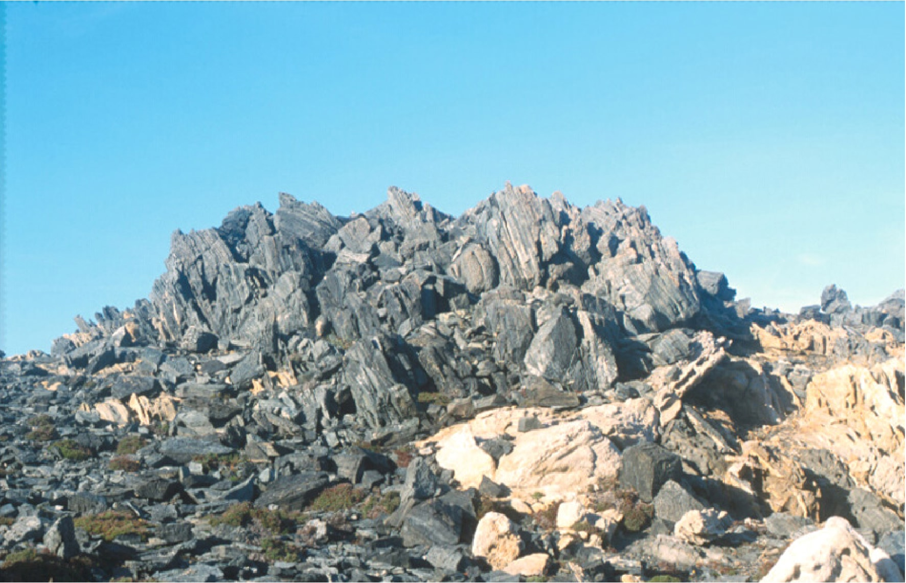
Following the road climbing toward the lighthouse we reach the northwestern slope of the promontory (Figure 2.9). Under the lighthouse, the diatexitic complex with granitoidic leucosomes involve relics of folded (F2) banded amphibolite and bodies of massive amphibolite and polyphase restitic bodies (Figure 2.10). Here, we can observe beautiful exposures of post-D2 high-temperature shear zones. The high-temperature shear zones seem to be involved by D3 upright mesoscale folds.
Figure 2.9. Panoramic view
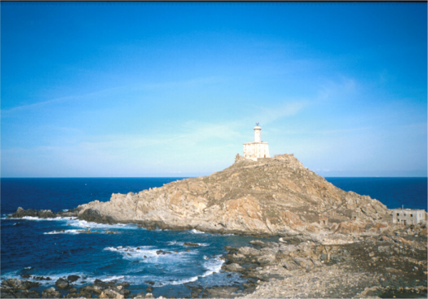
Panoramic view of the Punta Scorno Migmatitic Complex near the lighthouse (Stop 2.5).
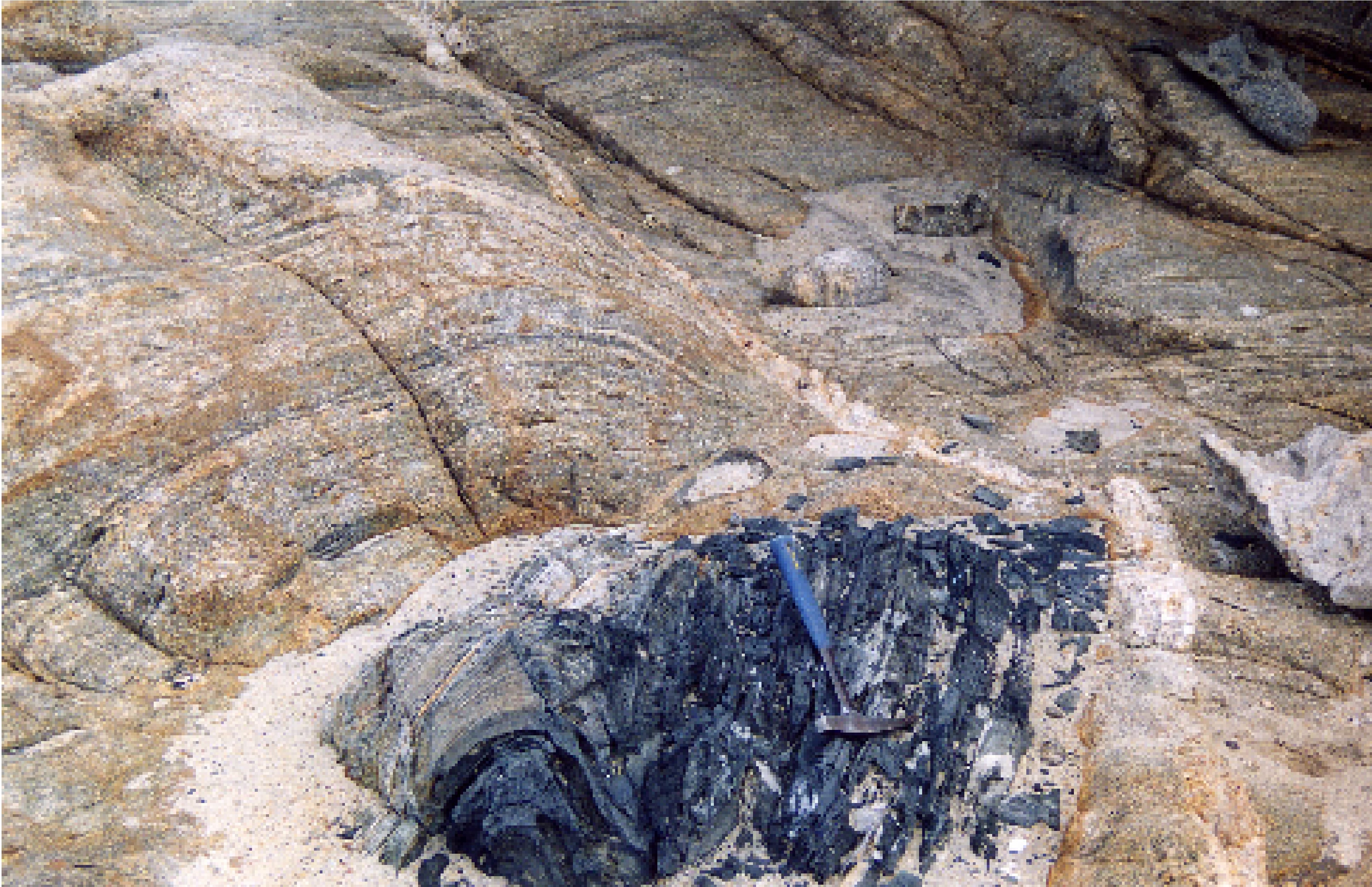
Figure 2.10. F2 fold in amphibolite

F2 fold in amphibolite embedded in the neosome component of the diatexitic complex. Punta Scorno area (Stop 2.5).
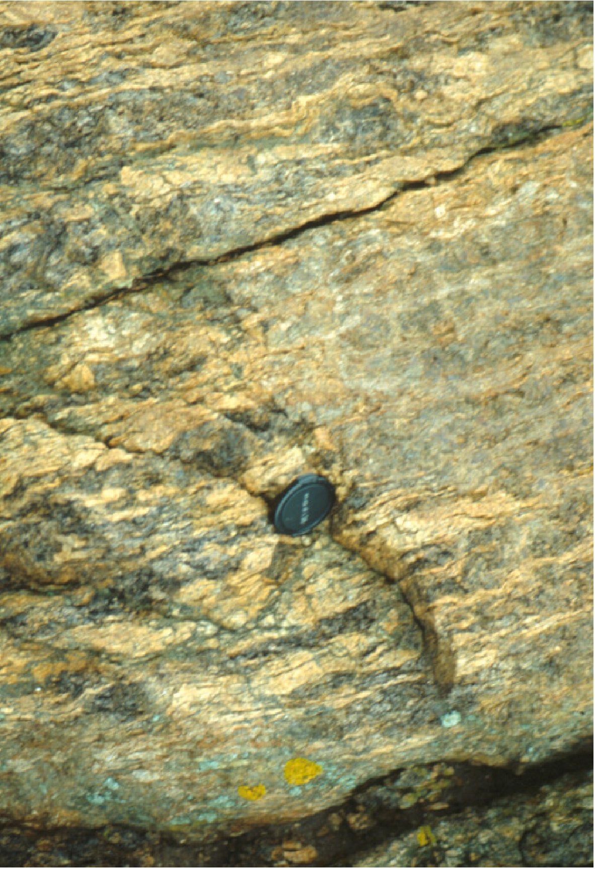
Rotated boudins of dark amphibolites of variable size (up to decametric-size) and leucocratic portions show a top-down to the north sense of shear, accommodating the late- or post-D2 exhumation of the HGMC (Figure 2.11A). Nice structures (probably formed under sub-magmatic flow condition) could be observed also in both the leucocratic and mesosome portions surrounding such sheared domains (Figure 2.11B) (Carosi et al., 2004a).
Figure 2.11. Sigmoidal structure in the leucosome
A. Sigmoidal structure in the leucosome provides a top-down-to-the-NE sense of shear in sections close to the XZ plane (Punta Scorno); B. Concordant and boudinated diffuse melanosome and pegmatitic dyke in diatexitic rocks. View orthogonal to the diatexitic foliation plane (Punta Scorno). Lens cap (52 mm) for scale. (Stop 2.5)
Itinerary 3: The Posada-Asinara Line in SW Gallura region
The third itinerary is focused on the transect, from the medium- to the high-grade metamorphic complexes, through the central sector of axial zone of the chain. We will focus on the regional systems of shear zones with opposite shear sense cropping out in the Giagazzu-Tungoni Complex (SW Gallura region) (Frassi, 2006). Few stops are dedicated to the migmatitic complex in the southern sector of Gallura region and on the metamorphic complex with a well-developed HT-LP metamorphic overprinting in Anglona region.
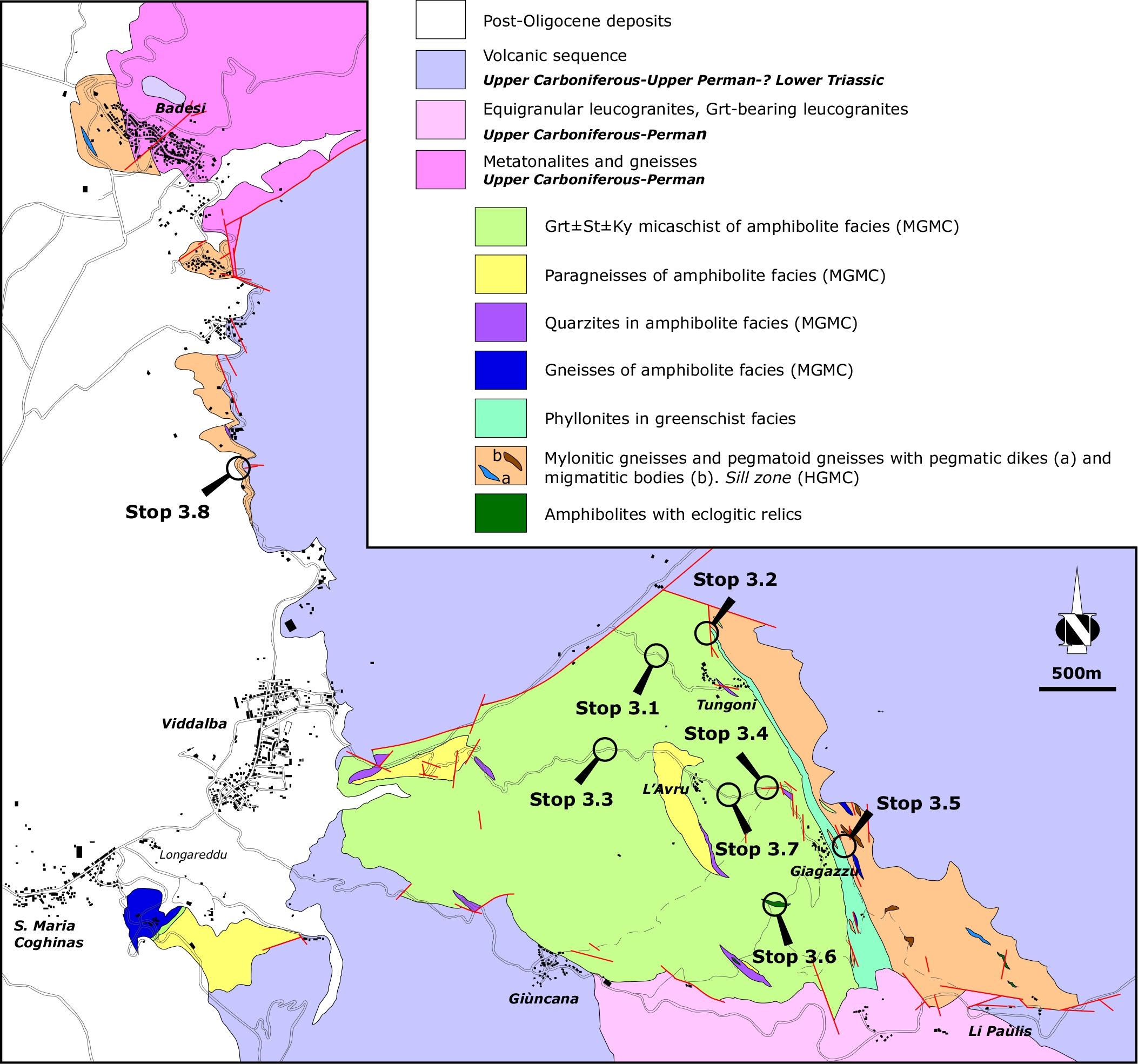
Figure 3.1. Geological map of the basement
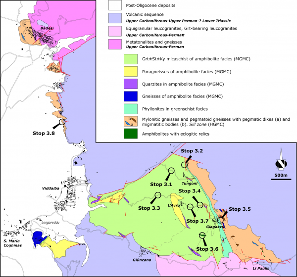
Geological map of the basement in the South Western Gallura and indication of the stops (modified after Frassi, 2006).
The northern sector of Posada-Asinara Line
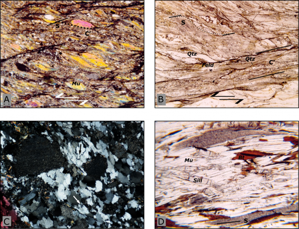
Stop 3.1- Along the road from Viddalba to Tempio Pausania villages, we turn on the second road on the right, toward the small village of Tungoni. After 300-350m the top-to-the SE shear zones are exposed in small outcrops on the left side of road. S-C and S-C’ structures develop on fine-grain size micaschists with muscovite, feldspar and quartz (Figure 3.2A) that present a quartz c-axis fabric intermediate between Type II and Type I crossed girdle (Figure 3.2B) (Frassi, 2006). The mylonitic foliation is the S2 foliation as highlighted by polygonal arcs and inclusion trails observed at microscopic scale.
Figure 3.2. The top-to-the SE shear zones
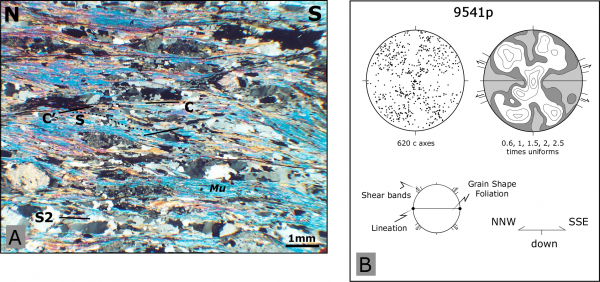
The top-to-the SE shear zones in the northern sector of Medium Grade Metamorphic Complex (Stop 3.1).
A. Photomicrograph of S-C' structures in the south of Tungoni village (crossed polarized light); B. Quartz c-axis fabric from continuous mm-thick quartz ribbons.
Stop 3.2- Turning on the right before the church of Tungoni, we can observe a continuous transect between the Ky-bearing mylonitic micaschists and the phyllonites.
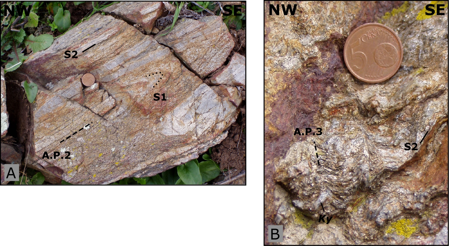
Walking 100-150 m outside the main street (Coord. N040° 55’ 31.6’’; E008° 56’ 34.4’’) we observe centimetric crystals of kyanite in levels and lenses developed parallel to the S2 mylonitic foliation. The kyanite is embedded by S2 foliation testifying its growth before the D2 deformation phase. Locally intrafoliar F2 folds (Figure 3.3A) and centimetric F3 folds have been preserved (Figure 3.3B) .
Figure 3.3. The Ky-bearing mylonitic micaschist (Stop 3.2).
A. F2 intrafoliar fold inside the S2 mylonitic foliation; B. Kyanite crystal enveloped by S2 mylonitic affected by centimetric F3 folds with sub-vertical axial plane.
Returning on the main road we can observe a gradual grain size reduction moving from the top-to-the SE shear zones (medium- grade metamorphic complex) toward the phyllonitic belt, where both S-C and S-C’ structures disappeared and the mylonitic foliation is highlighted by very fine-grained muscovite and small quartz ribbons (length < 1 mm) (Figure 3.4A).
The central sector of Posada-Asinara Line
Figure 3.4. Photomicrograph of phyllonites

Photomicrograph of phyllonites (left) (plane polarized light) (Stop 3.2) and Type 3 (Ramsay, 1967) F2-F3 fold interference pattern (right) in the Medium-Grade Metamorphic Complex near L'avru (Stop 3.3).
Coming back toward Viddalba we turn on the left to the small villages of L’avru and Giagazzu.
Stop 3.3- Near the L’avru village (Coord. N040° 54’ 54,5’’; E008° 56’ 12.7’’), type 3 (Ramsay, 1967) F2-F3 interference pattern can be observed (Figure 3.4B) because of the presence of centimetric and decimetric compositional layering that suggests a sedimentary origin for this complex. F2 folds show a chevron and/or kink geometry whereas the F3 folds are open folds with rounded hinges and sub-vertical axial planes.
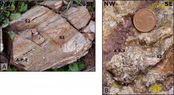
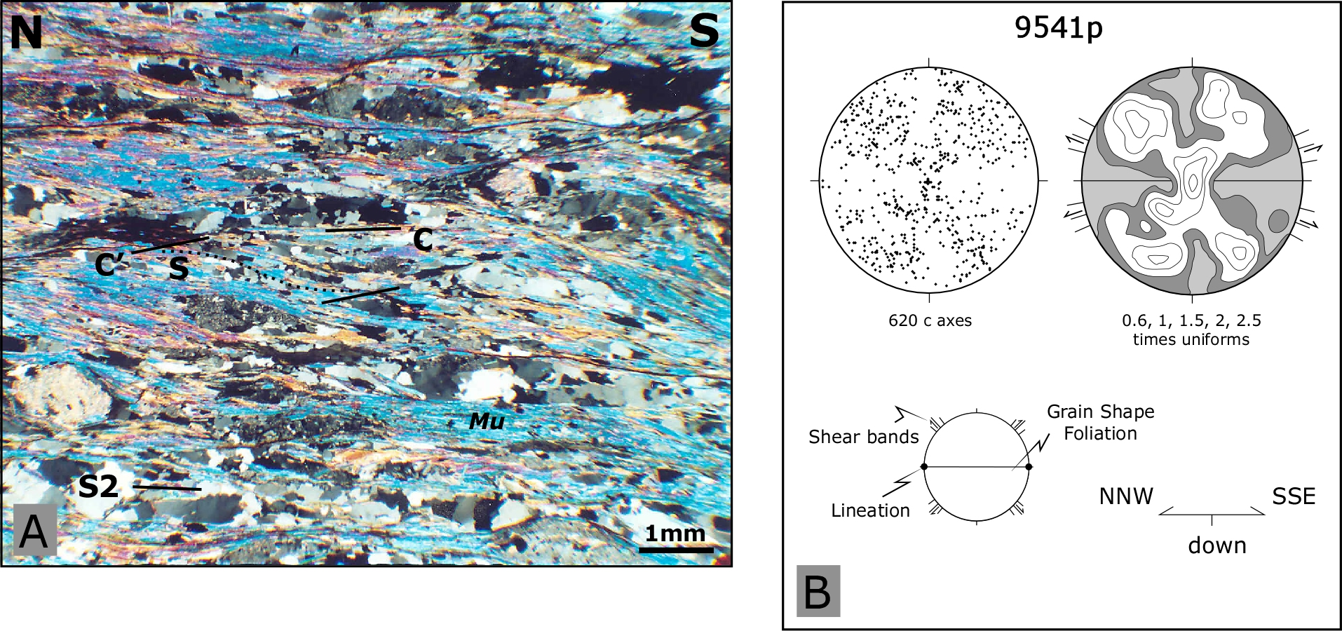
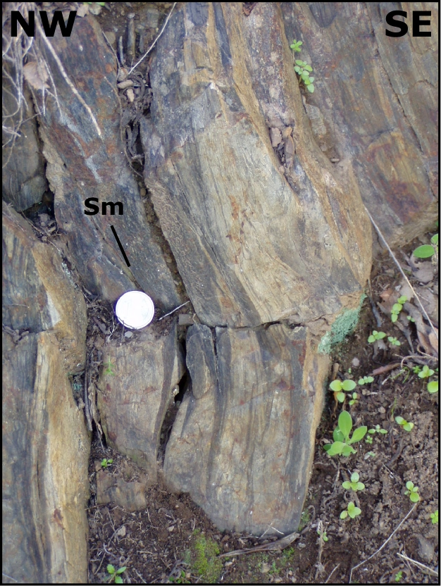
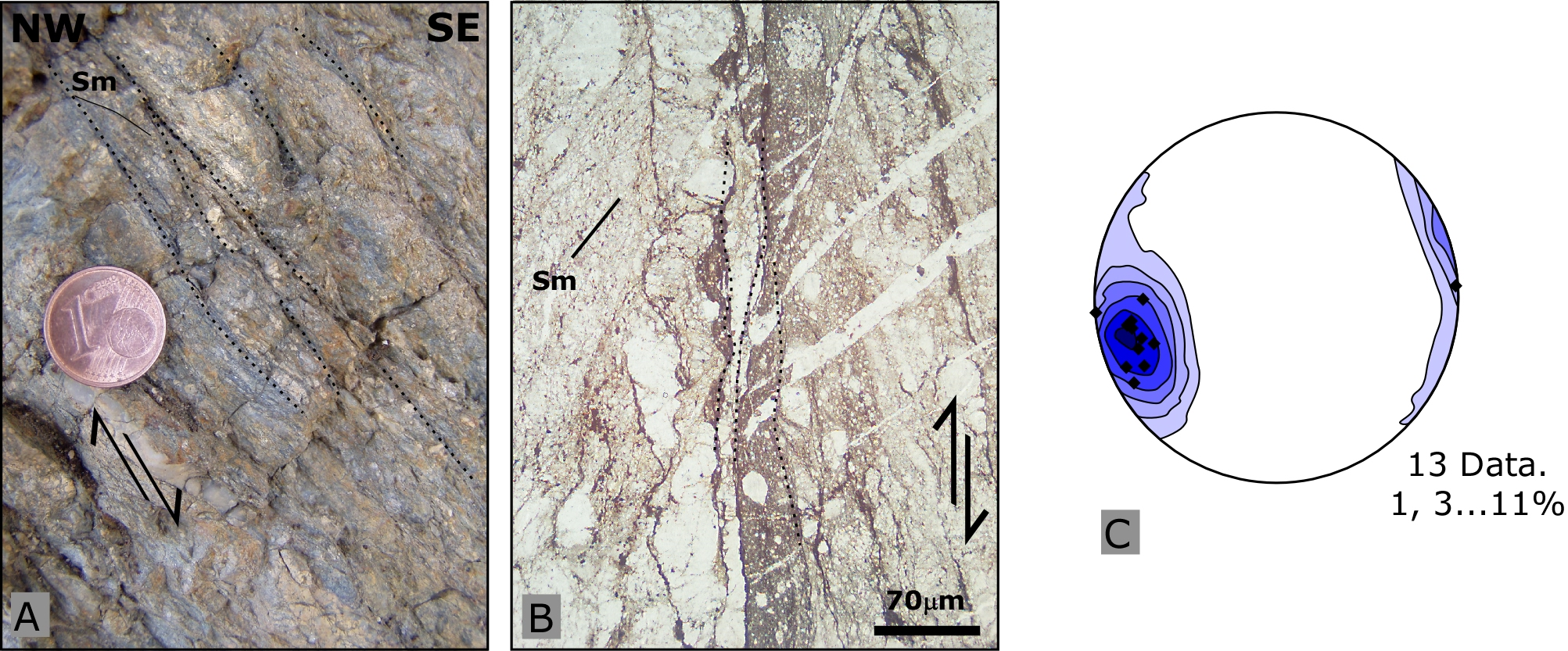
Stop 3.4- On the intersection for Giagazzu (Coord. N040° 54’ 42,3’’; E008° 57’ 00.6’’) we can observe the biggest outcrops (width ~100m) of top-to-the SE (“dextral”) shear zones in the MGMC. Mylonitic micaschists with cm-size crystals of kyanite and staurolite show a well-developed S-C’ and S-C structures clearly developed after the metamorphic peak (M1: Pl, Ky, St, Grt, Zo) (Figure 3.5A and B) . At the microscopic scale the mylonitic foliation Sm is marked by muscovite fishes, quartz ribbons, oxides, biotite, chlorite and quartz (Figure 3.5A and B) whereas the Chl+Rt association grown during the later stage of exhumation. The sigmoidal shape of quartz ribbons testifies a progressive transition from ductile to brittle-ductile deformation condition. The object lineation shows a NW-SE trend and a weak plunge toward NW.
Figure 3.5. The Ky±St±Grt bearing mylonitic micaschists near Giagazzu village (Stop 3.4).
A. and B. S-C and S-C' structures developed after the blastesis of Ky (kyanite), St (staurolite), Grt (garnet) and Pl (plagioclase) (Kretz, 1983). The mylonitic foliation is highlight by sigmoidal quartz ribbon, Mu-fish and asymmetric elongated crystal of plagioclase (A. plane polarized light; B. crossed polarized light); C. Quartz c-axis fabric from continuous mm-thick quartz ribbons.
Quartz c-axis measurements show a weak asymmetry, coherent with the top-to-the SE shearing highlighted at micro- and mesoscopic scale (Figure 3.5C) (Frassi, 2006).
Arrived at the Giagazzu church we walk about 650m across the phyllonites and the High Grade Metamorphic Complex.
Stop 3.5- Starting from Giagazzu small church (Coord. N040° 54’ 21,7’’; E008° 57’ 17.9’’) we follow a dirty road to Li Paùlis locality where we can observe the phyllonite belt and the deformation in the HGMC.
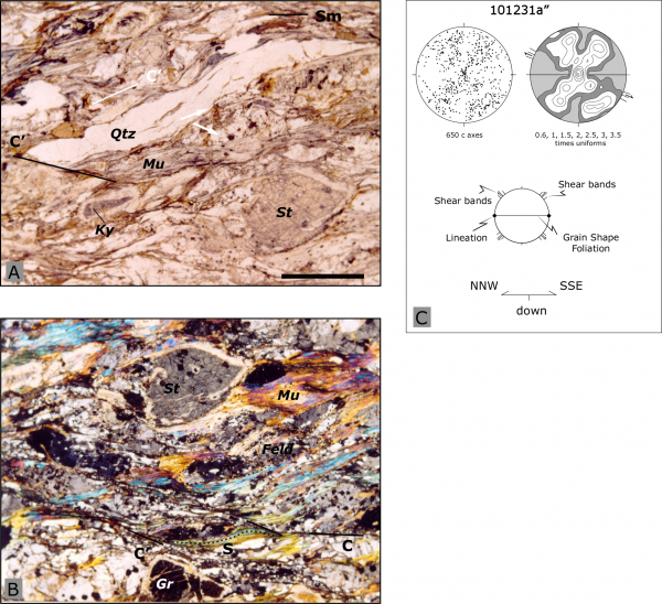
First of all we can observe the fine and homogenous grain size of phyllonites that here, are thicker then that in the Tungoni area (about 100m).
Rarely there are millimetric relics of feldspar and quartz limited in decimetric and centimetric levels parallel to the mylonitic foliation Sm. At microscopic scale these feldspar crystals (~20-30µm) show weak undulose extinction and rounded boundaries whereas quartz shows weak GBM and undulose extinction.
Near the last house on the left, we can observe an isoclinal F2 fold with the axial plane parallel to the Sm phyllonitic foliation (Figure 3.6), testifying the development of phyllonites during the compressive D2 deformation phase.
Going ahead the HGMC is defined by the continuous presence of porphyroblasts of feldspar inside a fine grain matrix organized in level parallel to the mylonitic foliation and rarely deeply replaced. The density and the grain size of feldspar grains in these levels vary from place to place and can be interpreted as relics of pegmatitic veins deformed in a sheared deformation regime. Metric migmatitic body with pegmatitic and/or leucosome veins can be locally observed.
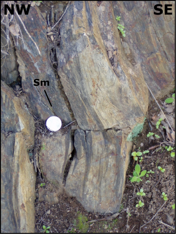
Frequently irregular networks of cm and mm black cataclasites with a top-to-the SE shear sense (Figure 3.7), cut the mylonitic foliation testifying that the dextral sense of movement was active until the upper structural levels, under brittle deformation conditions.
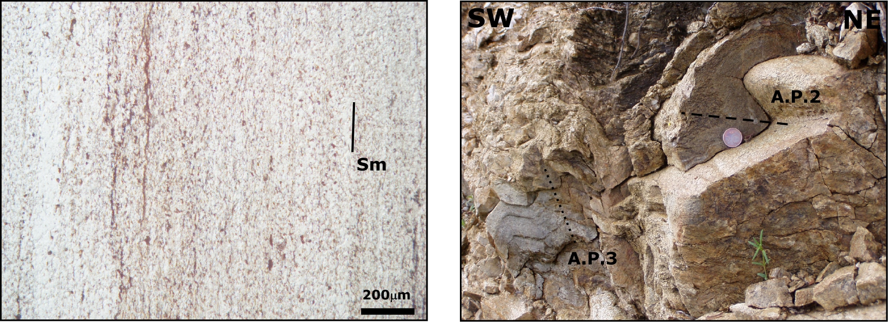
Figure 3.7. Brittle top-to-the SE shear zones in the Giagazzu-Tungoni
Brittle top-to-the SE shear zones in the Giagazzu-Tungoni in High Grade Metamorphic Complex (Stop 3.5).
A. Brittle shear zones oblique to the mylonitic foliation; B. Relationship between brittle top-to-the SE shear zones and Sm mylonitic foliation. Note the pressure solution on shear plane and the quartz veins apparently sheared with top-to-the NNW (PPL); C. Stereographic projection of cataclastic planes (Lower hemisphere, Schmidt projection).
It worth noting the presence, previously unrecognized, of top-to-the NW shear zones (“sinistral”) documented for the first time by Frassi (2006).
The biggest top-to-the NW shear zones outcrop is, along the same street, at about 600m from Giagazzu. The grain size is bigger than the previous HGMC outcrops. S-C and S-C’ structures, centimetric Mu-fish and sigma porphyroclasts of feldspar are well- developed (Figure 3.8A). Rarely centimetric quartz ribbons have been described (Figure 3.8B). Object lineation is highlighted by elongated quartz crystals and shows a NW-SE trend with weak plunge toward NW (SE).
Figure 3.8. Top-to-the NNW shear zones in the Giagazzu-Tungoni
Top-to-the NNW shear zones in the Giagazzu-Tungoni in High-Grade Metamorphic Complex (Stop 3.5).
A. and B. Examples of S-C' mylonitic foliation (CPL and PPL respectively); C. GBM and SGR in the relic quartz-feldspatic band preserved in the phyllonitic foliation (CPL); D. Sillimanite crystals (Sill), oblique to the mylonitic foliation Sm (PPL). Qtz- quartz; Mu-muscovite; Bt- biotite.
At microscopic scale the mylonitic foliation is highlighted by Mu-fish, by sigmoidal quartz ribbons and by elongated feldspars. Quartz shows well-developed undulose extinction, deformation bands, sub-grains and new-grains developed as consequence of GBM and weak SGR (Figure 3.9C). The feldspars show mechanical twinning locally interested by microfaults, and rare asymmetric myrmekites.
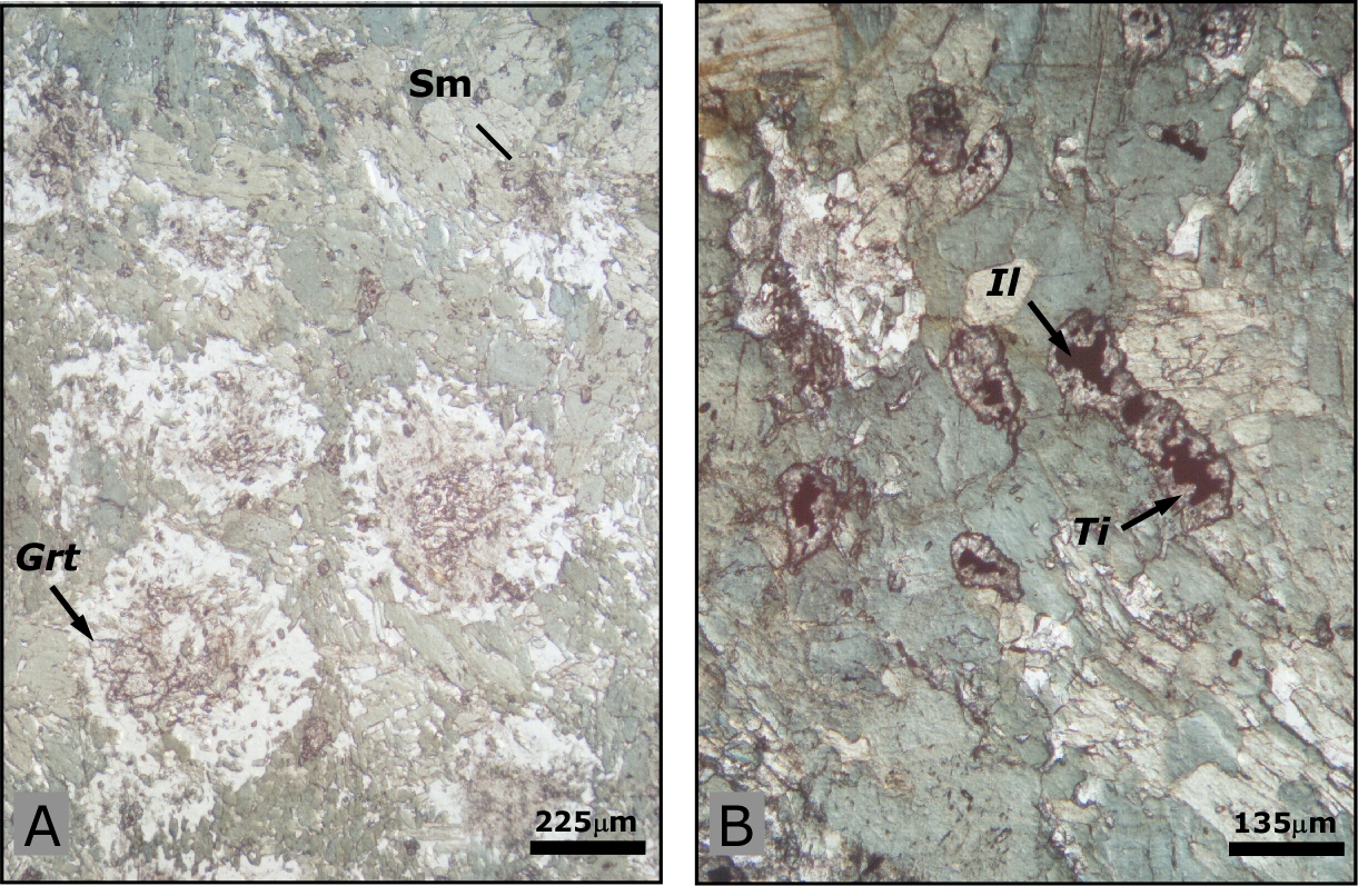
Figure 3.9. The retrogressed eclogitic body near Giagazzu village (Stop 3.6)
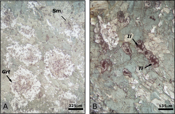
A. Photomicrograph of well preserved "eclogitic texture" with garnets (Grt) affected by fine grain symplectite aggregates of quartz, Ca amphibole and plagioclase (plane polarized light); B. Evidence of amphibolitic retrogressed event: ilmenite (Il) surrounded by titanite (Ti) rim.
Locally prismatic sillimanite growth parallel or/and oblique to the mylonitic foliation constrained its growth from early to late/post sinistral shearing (Figure 3.8D).
The retrogressed eclogite body in the MGMC
Stop 3.6- Within the micaschists, ~600m south-west of the Giagazzu village (Coord. N040° 54’ 07.2’’; E008° 56’ 59.4’’), an hectometric body of eclogite partially re-equilibrated under amphibolite facies occurs. This rock appears weakly deformed by the different deformation phases that affect the host micaschists. The texture is controlled by amphibole, garnet and plagioclase assemblage (Figure 3.9A). The evidence of amphibolitic retrogressed event are represented by the symplectites of clinopyroxene and albite, deriving from destabilization of omphacite, and by titanite rims on ilmenite crystals (Figure 3.9B).
The extensional tectonics
Stop 3.7- Coming back to L’avru, F5 fold can be observed. This system of folds shows an asymmetric geometry and a sub- horizontal axial plane. For these reasons they could be interpreted as consequence of gravity collapse developed in an extensional regime during the later tectonic stages.
The top-to-the NW mylonitic belt in the Badesi Complex
Returning to Viddalba village we turn on the right on the road to Badesi village.
Stop 3.8- Before the Azzagulta village we can observe, in a little quarry (Coord. N040° 56’ 11,4’’; E008° 53’ 32.0’’), the biggest outcrop of sinistral shear sense mylonites. Very elongated quartz ribbons, often around feldspars, S-C and S-C’ structures and sigma and delta porphyroclasts are well developed.
The object lineation is represented by Qtz-ribbons, elonged Qtz-Pl aggregates and by micro-faulting and micro-boudinage of feldspars.
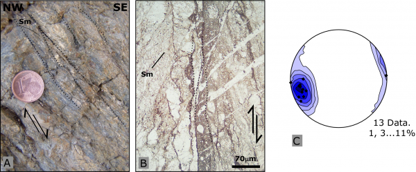
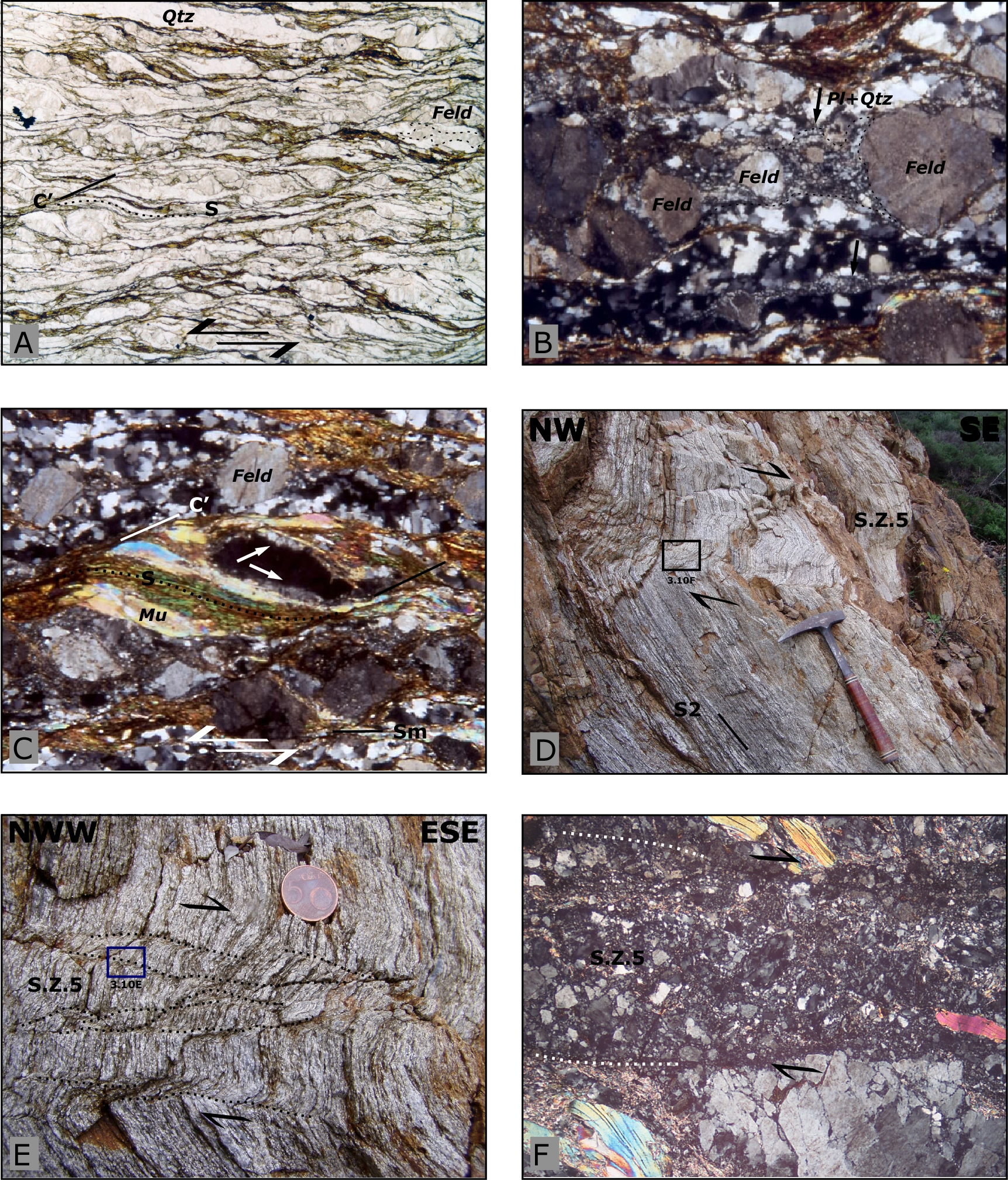
At microscopic scale the C’ planes are highlighted mostly by oxides and little crystals of muscovite, biotite and quartz (Figure 3.10A) locally completely recrystallized (Regime II-III, Hirth & Tullis, 1992). The mylonitic foliation Sm, is highlighted by Mu-fish, elongated feldspar, often completely replaced by myrmekites (Figure 3.10B) and by sigmoidal quartz ribbons (Figure 3.10A). Delta porphyroclasts of feldspar, muscovite fishes and asymmetryc myrmekytes are the main kinematic indicators (Figure 3.10C). Rare fibrolite has been described.
Figure 3.10. Top-to-the NW mylonitic belt in the Badesi Complex (Stop 3.8).
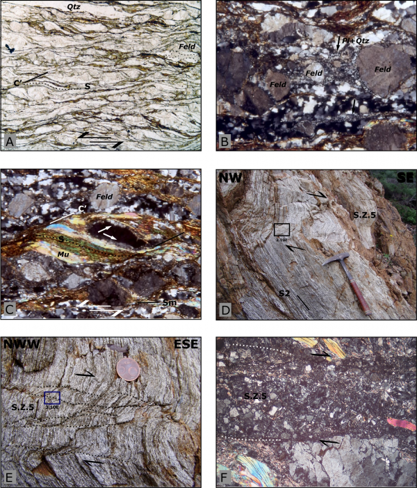
A. Sigmoidal quartz ribbon in S-C' structures (CPL); B. Qtz-Pl microcrystalline ribbons (arrows) as consequences of myrmekites development (CPL); C. Asymmetric myrmekites developed in the strain caps of feldspar crystal (CPL);
D. and E. Extensional brittle S.Z.5 shear zones developed on the F5 vertical limbs in the Badesi Complex; F. Photomicrograph of top-to-the SE brittle S.Z.5 shear zone (CPL).
Where the mylonitic foliation present sub-vertical dip, brittle-ductile and brittle shear zones have been developed as axial planes of metric and centimetric gravity collapse folds (Figure 3.10D, E and F).
The quartz c-axis CPO analyses carried out on sample caming from this mylonitic complex show locally a well developed asymmetric fabric in agreement with the shear sense indicators at mesoscopic scale. The geometry is, generally, close to plane strain (Frassi, 2006).
Samples coming from this outcrop show vorticity numbers (Wm) ranging from 0.80 and 0.88, highlight a predominant simple shear component deformation (Frassi, 2006).
Itinerary 4: Northeastern Sardinia
We move to the northeastern coast of Sardinia and we cross the Variscan structure from north to south starting from the High-Grade Metamorphic Complex to reach the Medium-Grade Metamorphic Complex NE of Mt. Albo.
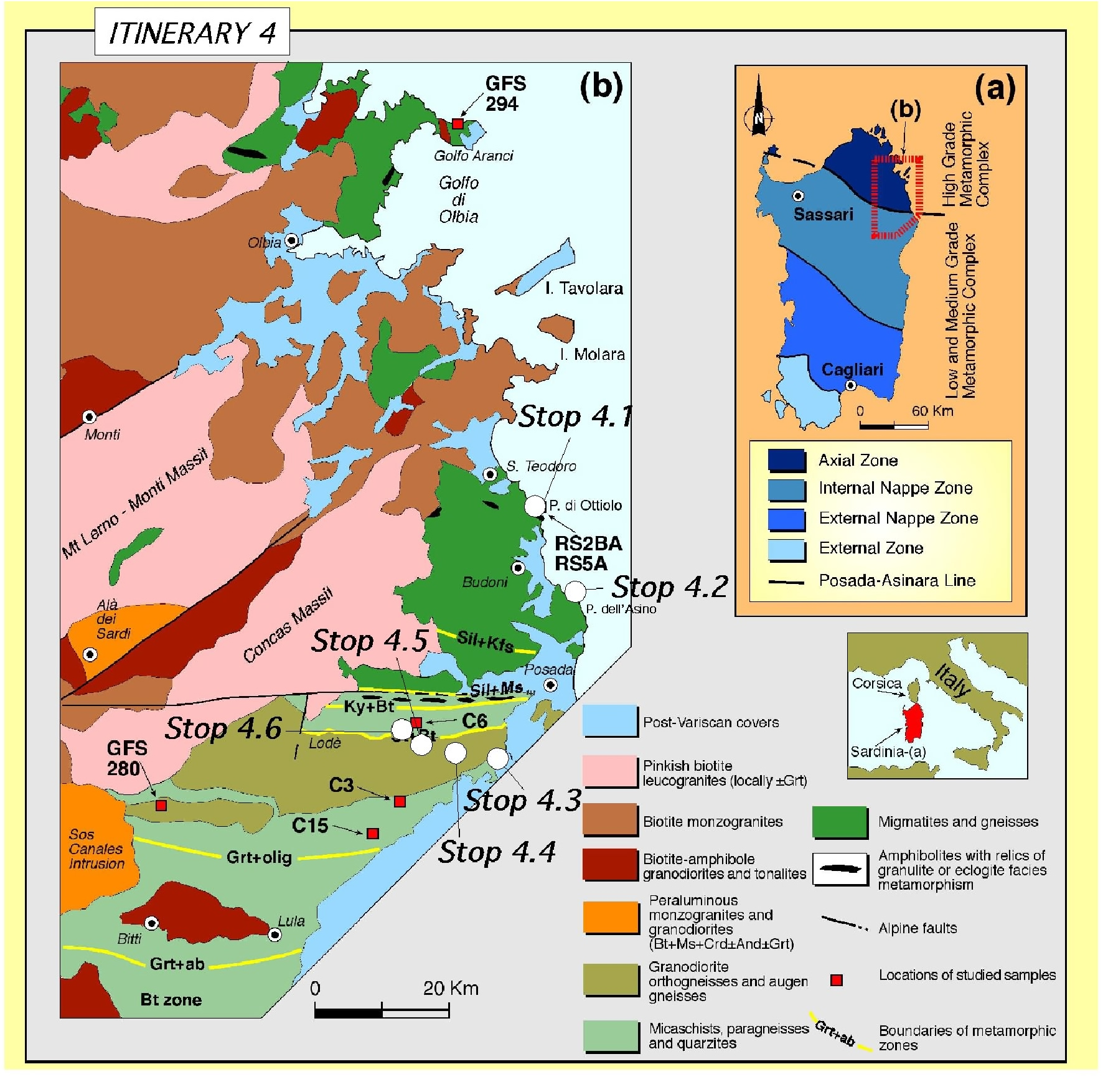
This itinerary (Figure 4.1)(Figure 4.2)(Figure 4.3a and b) has been modified after the field guide books of Carmignani etal., 1982 (Società Geologica Italiana), 1986 (IGCP project n.5), 1993 (Gruppo Informale di Geologia Struttuarle), 1994 (16th General Meeting of the International Mineralogical Association) and review by Franceschelli et al. (2005).

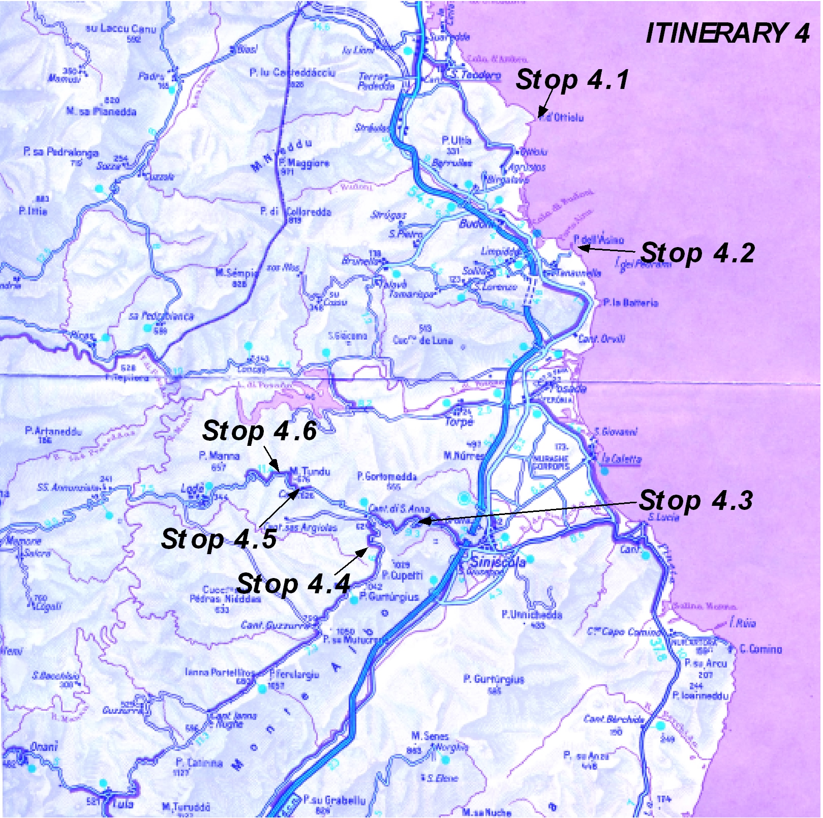
Figure 4.2. Stops of the itinerary in the Baronie region
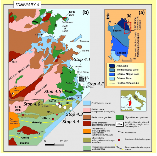
Stops of the itinerary in the Baronie region (modified after Di Vincenzo et al., 2004).
Figure 4.3. N-S geological cross section in the Posada Valley
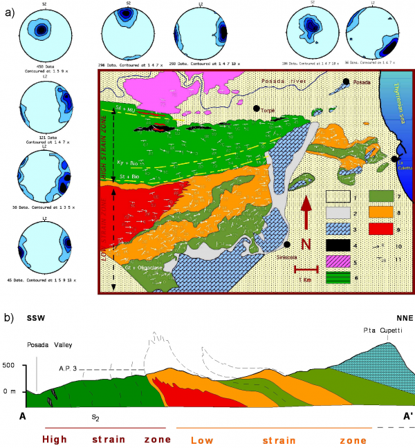
a) See the geological map (Figure 4.3B) for explanations of the colours (modified after Carosi & Palmeri, 2002).
b) Geological and structural map of the Baronie area south of Posada Valley (modified after Carosi & Palmeri, 2002).
b) 1: Continental beach deposits (Pliocene-Quaternary); 2: Slope debris (Quaternary); 3: Carbonate platform sediments, continental and evaporites (Triassic-Jurassic-Cretaceous); 4: Amphibolites with relics of granulite facies parageneses; 5: Migmatitic complex of Sill + Mu and Sill + K-feldspar zones (344+/-7 Ma); 6: Micaschists and porphyroblstic paragneisses of Stu + Bt and Ky + Bt zones; 7: Phyllites and metasandstones of Bt zone; micaschists and paragneisses of Grt + Ab and Grt + (Albite+Oligoclase) zones; 8: Granitic augen gneisses (441+/-33Ma); 9: Granodioritc orthogneisses (458+/-31 Ma); 10: trend and plunge of L2 stretching lineation; 11: strike and dip of mylonitic foliation (S2).
We reach Olbia and we continue to the South along the SS125 road “Orientale Sarda”. The first stop is on the left in correspondence of the turistic village of Porto Ottiolu.
Stop 4.1- Porto Ottiolu - Punta de li Tulchi.
From Porto Ottiolu we follow a footpath for few hundred of metres going to Punta de Li Tulchi where we can observe migmatites of sillimanite + k-feldspar zone, calc-silicate nodules and boudins of retrogressed eclogites.
The migmatites are constituted by migmatized orthogneisses, biotite-rich gneisses, stromatic migmatites. At Punta Ottiolu stromatic migmatites, dictyonitic and nebulitic migmatites and discordant decimetric leucosomes crop out. Dyctionitic leucosomes, fine grained and with granitic composition, are abundant within the shear bands of the orthogneiss.
Centimetric-size shear bands, related to emplacement of leucosomes, affect the migmatites. They have been interpreted by Elter et al., (1999) as a regional system of shear zones affecting the migmatites during collision, connected to a network of extensional shear zones giving rise to an extensional gneiss dome in the HGMC of NE Sardinia.
According to Carmignani et al., (1994b) we can observe a considerable evolution of the leucosomes at the point that the stripped or banded structures disappear and the rock loses the regular layering. In the migmatites we can found gray-green calc-silicate nodules and pods (5-30 cm). They have spheroidal to ellipsoidal shapes, are fine grained and lack in any foliation.
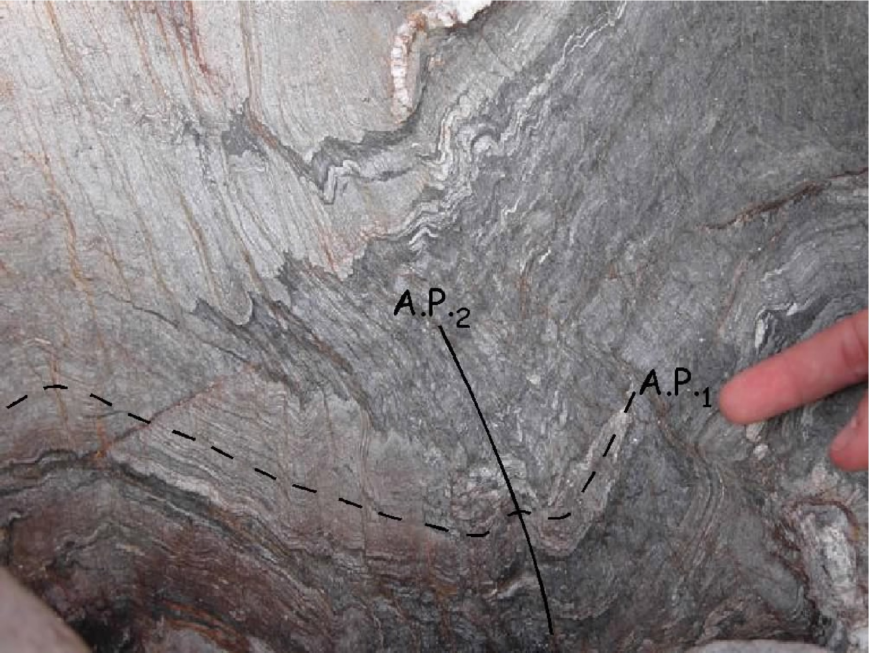
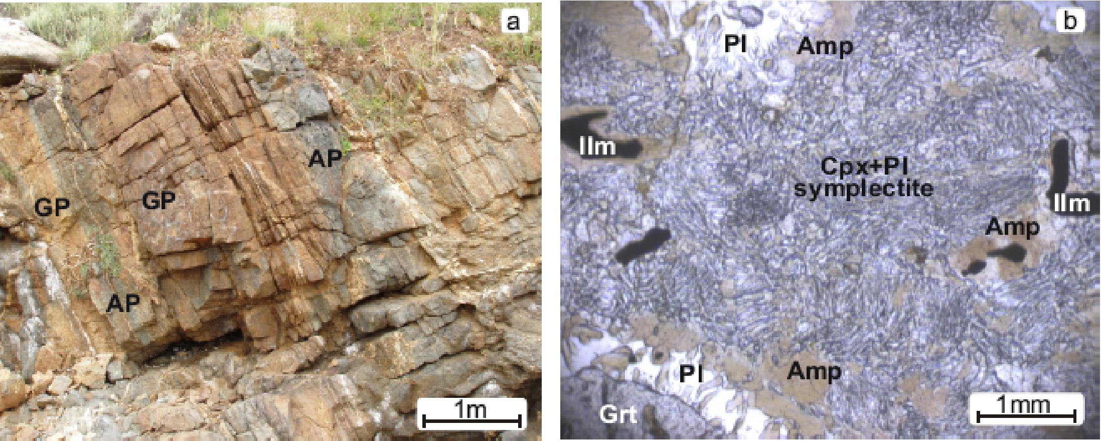
According to Franceschelli et al., (1998) two main types of metabasites with eclogite relics can be distinguished: garnet- pyroxene-rich and amphibole-plagioclase-rich layers (Figure 4.4).
Figure 4.4. The metabasites with eclogite (Stop 4.1).

A. Alternation of garnet-pyroxene (GP) layers and amphibole-plagioclase (AP) layers in the eclogite outcrop at Punta de li Tulchi (NE Sardinia). Photomicrographs showing metamorphic evolution of eclogite from Punta de li Tulchi; B. Diopsidic clinopyroxene and Na-plagioclase symplectite resulting from destabilisation of omphacites; in the upper and lower left corners, note the kelyphytic structure around garnet. One polar (from Franceschelli et al., 2005).
The meaning and the age of the eclogites is still controversial and debated since the paper by Cappelli et al., (1992) interpreting them as an evidence of a paleosuture and giving to the protolithes an age of 957±93 Ma. Recent U-Pb zircon data point to protolith ages at around 460 Ma (Cortesogno et al., 2004; Palmeri et al., 2004; Giacomini et al., 2005).
We come back to the SS125 road “Orientale sarda” and we continue southward to the crossroad to the village of Tanaunella. We move few hundred meters northward to Porto Ainu and we walk a little bit to reach Punta dell’Asino.
Stop 4.2- Punta Batteria – Punta dell’Asino. Migmatites and associated rocks of sillimanite+k-feldspar zone. (Coord. N40° 41’ 34.7’’ ; E 009° 44’ 12.5’’).
A typical sequence of the migmatitic complex of the sillimanite+k-feldspar is exposed along the coast between Punta Battera and Punta dell’Asino. Moving to the north we encounter different lithologic types such as:
- biotite-sillimanite mesocratic gneisses (Figure 4.5);
- stromatic migmatites with rare Ca-silicate lenses;
- stromatic migmatites with abundant leucosomes and mesocratic gneisses and Ca-silicate lenses and nodules;
- granodioritic orthogneisses.
The main foliation is the regional S2 foliation striking about E-W and dipping towards the south. Mesoscopic evidences of pre-S2 foliation are scanty; late folds with variable geometries are abundant (Figure 4.6).

Biotite-sillimanite mesocratic gneisses in the outcrop of Punta dell’Asino show, on the foliation, abundant iso-oriented rods constituted by quartz and fibrolitic sillimanite.
According to Franceschelli et al., (1991) they are the result of the decomposition of biotite induced by the activity of hydrogen ions through the model reation:
Bt+H+=Sil+SiO2+(K+,Mg2+, Fe2+)+H2O.
Leucosomes are rare in this outcrop whereas they are abundant near Mt. Rasu: here the rock bodies can reach up to 1m in thickness. The leucosomes are sometimes garnetiferous with aplogranite composition and parageneses which include Qtz+Pl+Kfs+Grt±Bt±Sil± Ms. Associated mesocratic gneisses are lacking in k-feldspar and contain, together with Qtz+Pl+Bt+Ms+Grt+Sil, some relics of kyanite.
Near Mt. Ruju, mesocratic gneisses are partially lacking in leucosomes, and contain abundant Ca-silicates nodules. These nodules have spheroidal to ellipsoidal shapes, are fine grained, and lack in any foliation. They show concentric zoning: the light green to pink core consists of Qtz+Pl+Cpx+Grt; the green to dark green rim is characterized by the increase of the amount of horneblende and sphene, instead of Cpx+Grt.
The granodioritic orthogneiss crops out near Mt. Nuraghe. It shows augen to banded structures and is relatively homogeneous, containing only some E-W trending mafic inclusions.
Based both on petrographic and chemical data, the orthogneiss is quite similar to the granodioritic orthogneiss of Lodè and plot on the same Rb-Sr whole rock isochron of 458±31 Ma (Ferrara et al., 1978). New U-Pb analyses on zircons confirm this age (458±7 Ma; Helbing & Tiepolo, 2005).
We come back to the road SS125 "Orientale Sarda" road and we continue southward to Siniscola and then to Lodè villages.
Stop 4.3- Road from Siniscola to Cantoniera di S. Anna and Lodè. Granitic augen gneiss (coord. N40° 35’ 00.7’’; E 008° 39’ 47.4’’).
We stop on the hairpin bends of the road few kilometres before reaching the upland, to observe granitic augen gneiss deformed by the D2 shearing phase and showing shear bands (Figure 4.7). The granitic augen gneiss is mainly made up of layered bodies of augen gneisses alternated with thinner micaschist levels. The rocks have been originally considered the product of Variscan metamorphism over rhyolites and arkosic sandstone (Ferrara et al., 1978) but they could represent an intrusive facies of Ordovician granitoids. The age has been constrained at 441±33 Ma (Rb-Sr whole rock; Ferrara et al., 1978).

Stop 4.4- Road from Siniscola - Cantoniera di S. Anna - Lodè. (Coord. N40° 04’ 20.0’’; E 009° 38’ 11.6’’).
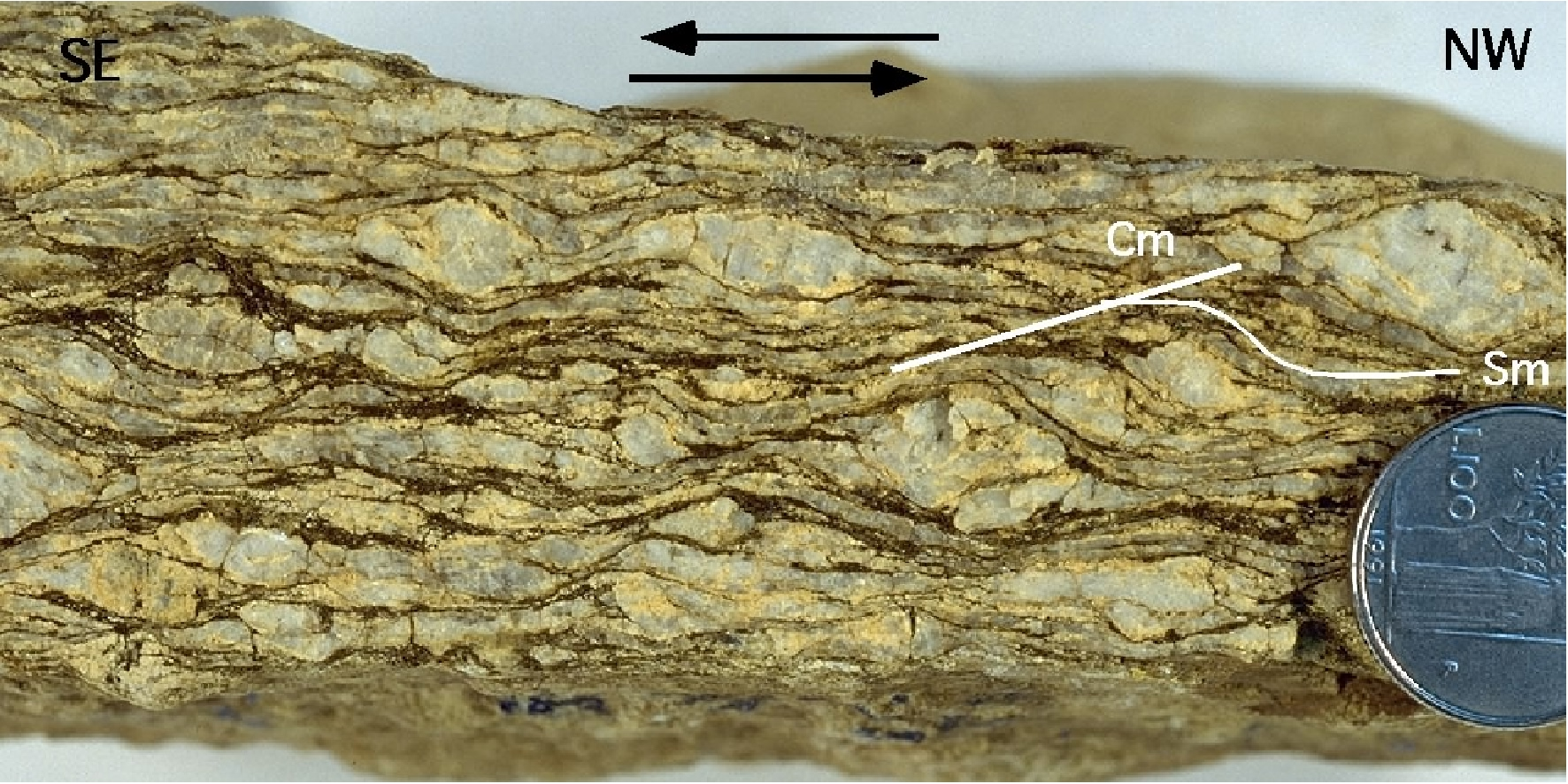
At Cantoniera di S. Anna we turn on the left on the road to Lula village and on the northeastern slope of Mt. Albo (made up of mesozoic limestones, dipping to the SE) we can observe the metamorphites of garnet + albite + oligoclase zone: micaschists, porphyroblastic paragneisses (Figure 4.8) and granitic augen gneisses (Figure 4.7).
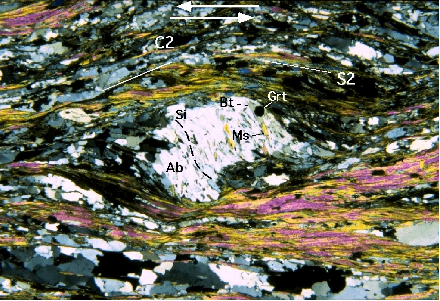
The contact between the basal levels of the porphyroblastic paragneisses and the granitic augen gneisses is visible. It is the southern limb of the Mamone-Siniscola D2 antiform (Figure 4.3a and b): its nearly E-W trending axis, plunging to the est. The porphyroblastic paragneisses are characterized by the occurrence of millimetric plagioclase porphyroblasts with albite core and oligoclase rim (Franceschelli et al., 1982) showing evidence of post-D1 and pre-D2 growth and includng an internal foliation (S1). Internal S1 foliation is defined by inclusion trails of white mica, quartz, garnet and minor biotite (Figure 4.9) and (Figure 4.10). Syn-D1 white mica are invariably celadonite-rich and paragonite-poor, whereas D2 micas usually show low-celadonite and high-paragonite composition. The external foliation envelops porphyroblasts and it is an advanced S2 crenulation cleavage dominated by white mica and minor chlorite and biotite. Garnet, oligoclase and opaque minerals are also found along S2. Microlithons relics of the S1 foliation and F1 fold hinges are preserved within S2.
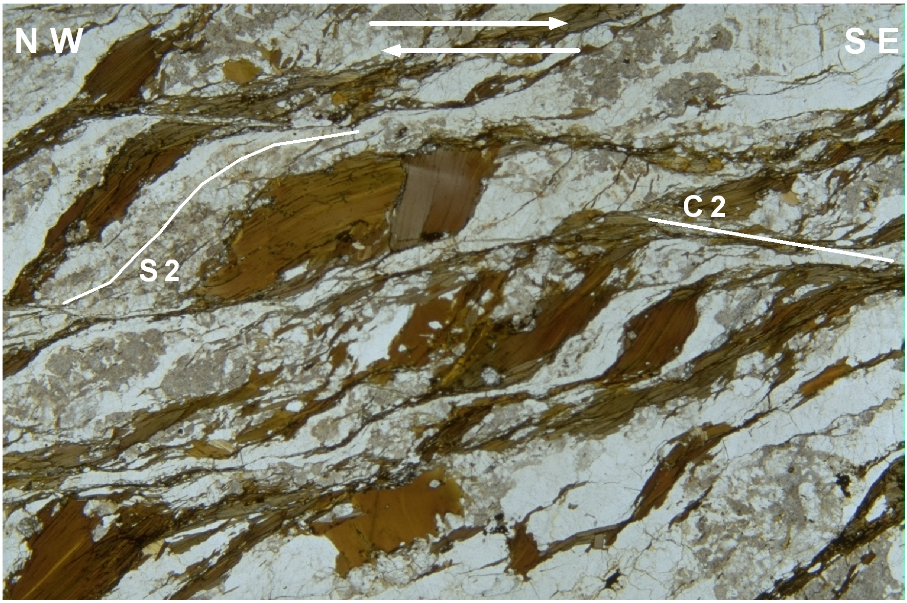
Figure 4.9. Albite porhyroblast
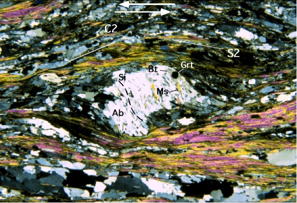
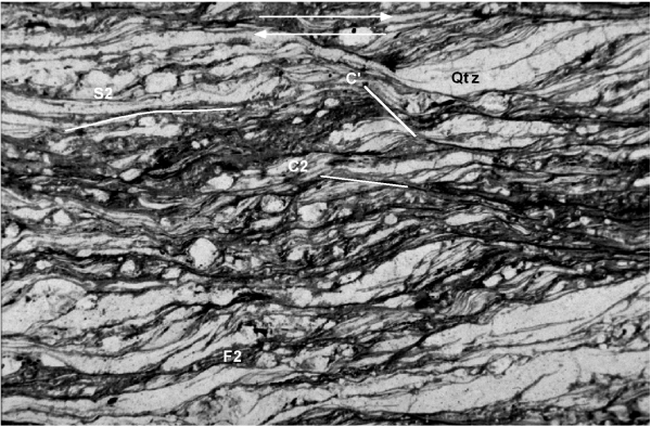
Albite porhyroblast (Ab) including a sigmoidal inclusion pattern (Si) marked by quartz, garnet (Grt), biotite (Bt), muscovite (Ms) and Graphite (Stop 4.4). Si is nearly continuous with S2 foliation. S2-C2 fabric point to a top-to NW sense of shear (Micaschist of the garnet + (albite+oligoclase) zone, CPL, 52x) (Carosi & Palmeri, 2002).
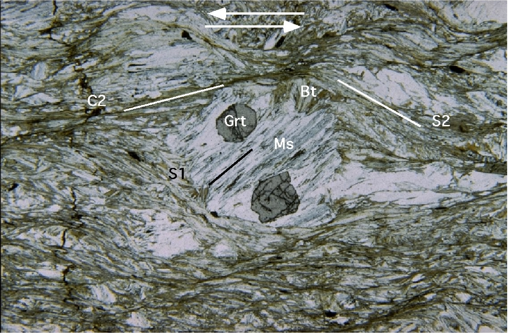
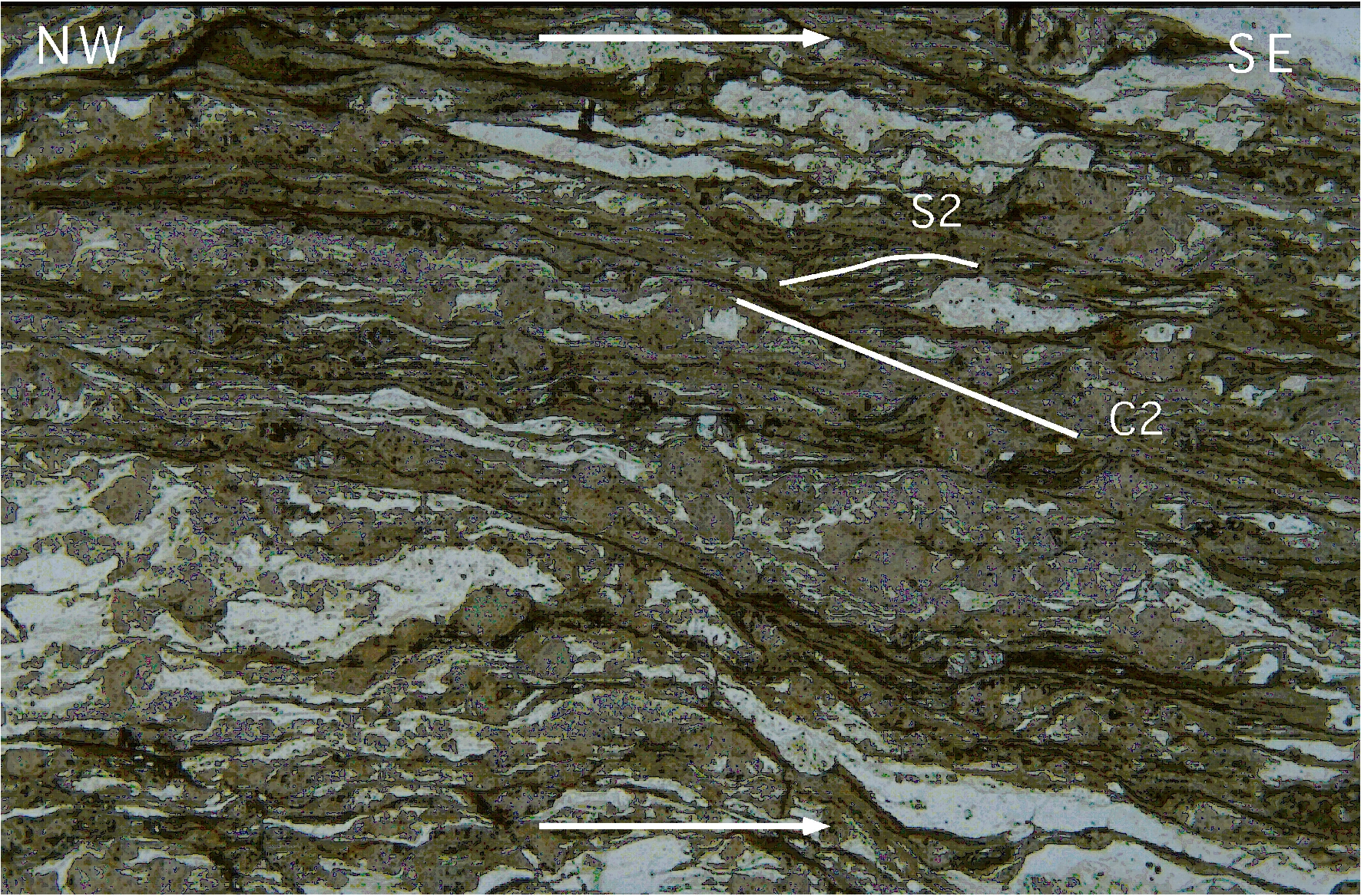
Figure 4.10. Albite porhyroblast

Albite porhyroblast including a straight inclusion pattern (S1 foliation), marked by garnet (Grt), biotite (Bt), muscovite (Ms) and graphite (Stop 4.4). S2-C2 fabric point to a top-to NW sense of shear (Micaschists of the Garnet + (Albite+Oligoclase) zone, PPL, 52 x).
In situ Ar-Ar laser analyses of white micas yelded ages of ~ 340-315 Ma (Di Vincenzo et al., 2004). It is worth noting that the oldest ages (335-340 Ma) were detected in syn-D1 white mica generation not texturally and chemically re-equilibrated at upper crustal levels. Syn-D2 white mica ages cluster at 315-320 Ma.
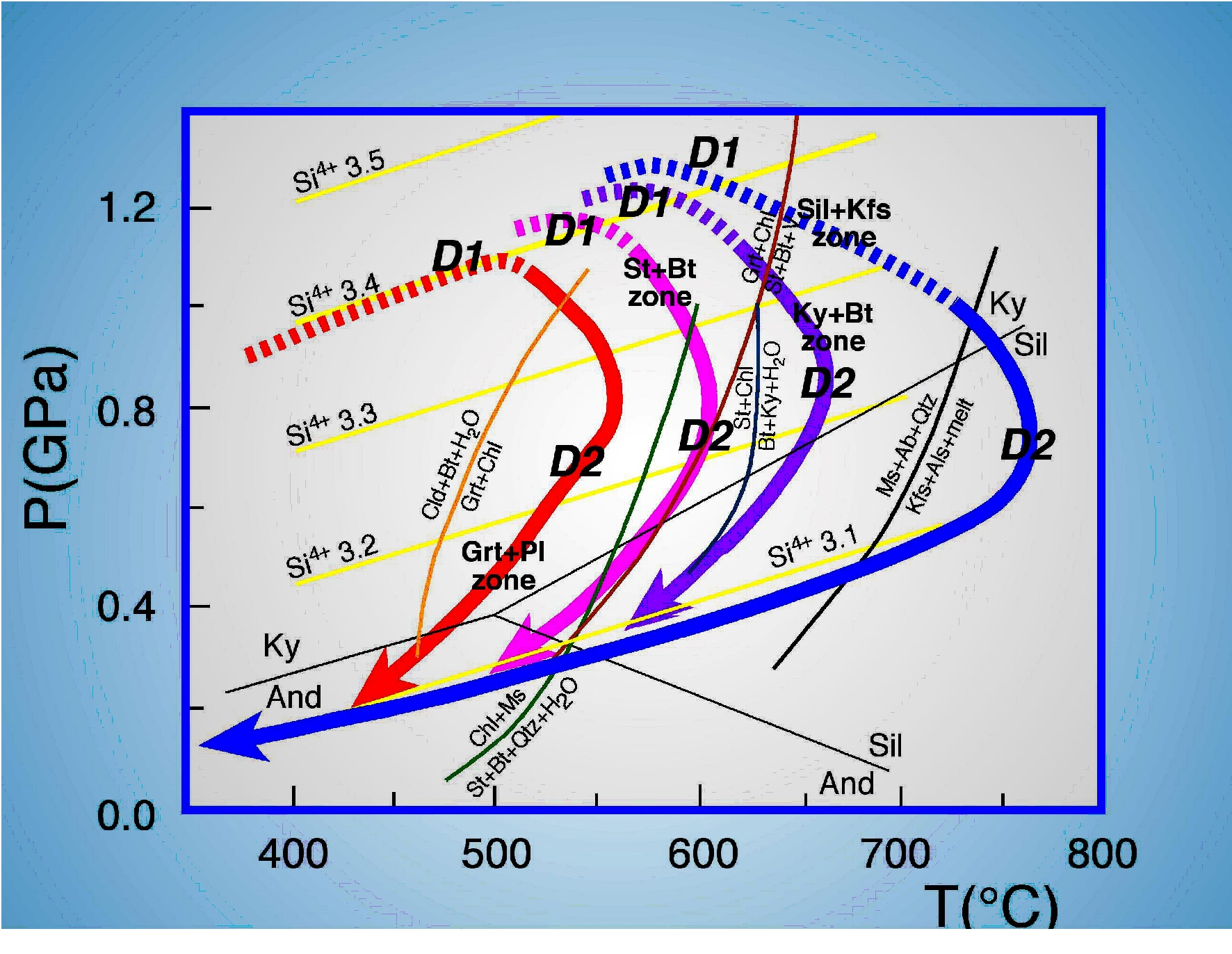
The estimated temperatures for the garnet-albite-oligoclase zone range from 453°C to 521°C moving from the upper to the deeper part of this zone. Pressure obtained form Grt-Bt-Ms were around 0.7-0.8 GPa (Franceschelli et al., 1989). Carosi & Palmeri, (2002) and Di Vincenzo et al., (2004) reported temperatures of 500-550°C and pressures of 0.8-1.1 GPa during D1 and 550-600°C and pressures 0.7-0.9 GPa for the D2 phase (Figure 4.11) and (Figure 4.12).
Figure 4.11. P-T-t paths
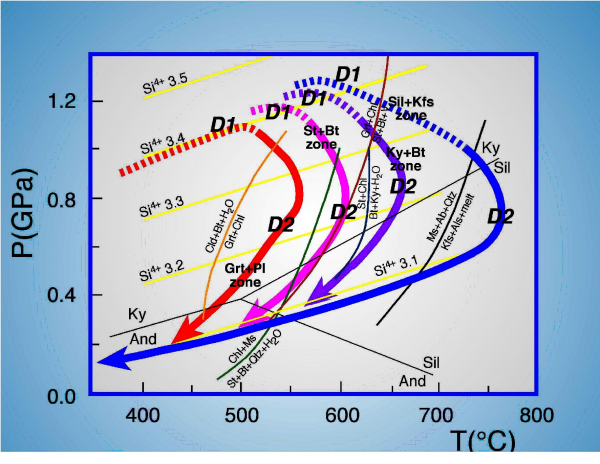
P-T-t paths for the different metamorphic zones of NE Sardinia (modified after Di Vincenzo et al., 2004)(Stop 4.4).
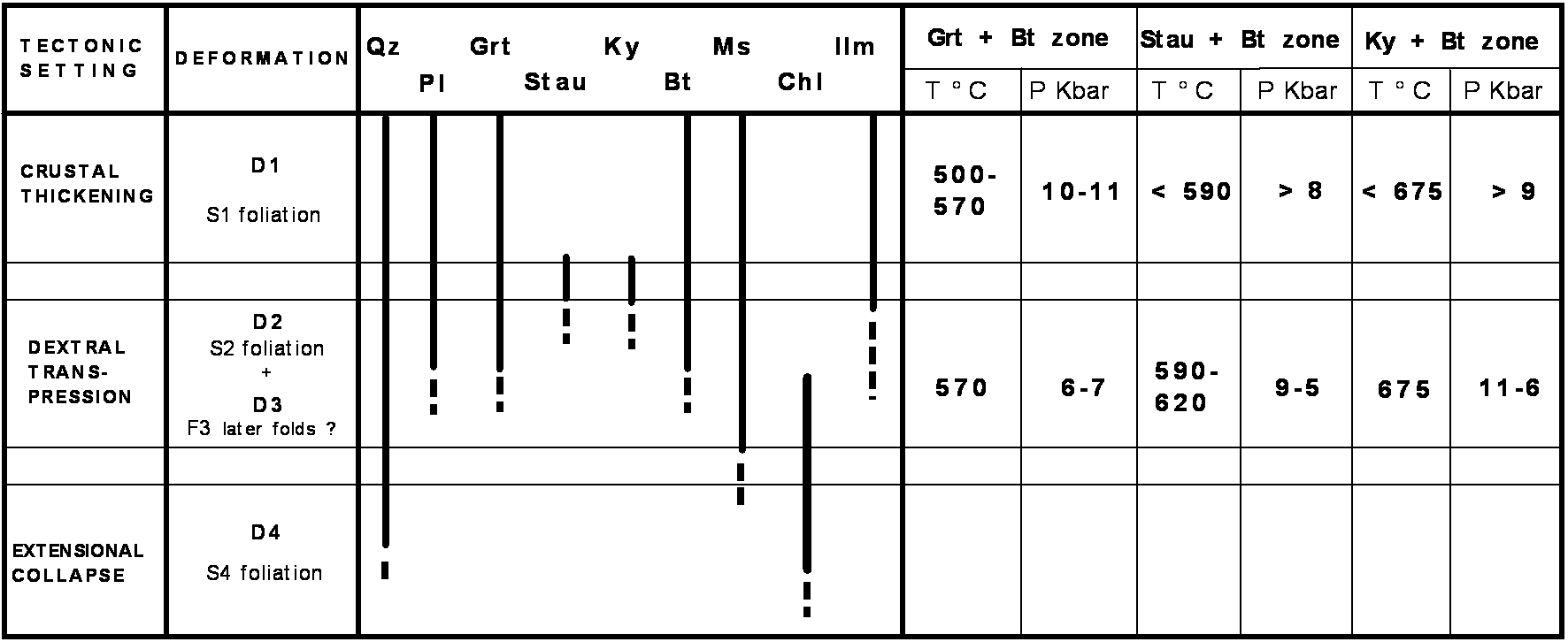
Figure 4.12. Synoptic table

Synoptic table of the deformation phases and related estimations of pressures (kbar) and temperatures (°C) (Carosi & Palmeri, 2002) (Stop 4.4).
Stop 4.5- Road from Siniscola to Lodè. Cantoniera Mt. Tundu. Granodioritic orthogneiss with C-S fabric. (Coord. N40° 35’ 40.4’’; E009° 35’ 34.0’’).
Few kilometers after Cantoniera di S. Anna we stop on the right at Cantoniera di Mt. Tundu. Few dozen metres north of the Cantoniera house we can observe granodioritic orthogneiss with a prominent C-S fabric and C’ structures pointing to a top-to-the NW sense of shear ("dextral")(Figure 4.13) and (Figure 4.14). The main foliation (S2) is nearly vertical and strikes nearly E-W and preserve a large number of melanocratic inclusions which are flattened along the S2 foliation.
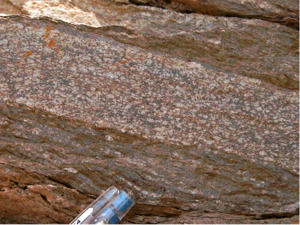
Figure 4.13. C-S fabric

C-S fabric in the granodioritic orthogneiss (top-to-the NW sense of shear) (Stop 4.5).
Figure 4.14. Photomicrograph of biotite fish

Photomicrograph of biotite fish in mylonitic granodioritic orthogneisses (top-to-the SE sense of shear. PPL; fov is nearly 3 mm) (Stop 4.5).
In origin they were intrusive rocks of granodioritic composition with a radiometric age of 458±31 Ma (RbSr whole rock; Ferrara et al., 1978) and 456±33 Ma (U-Pb on zircons; Helbing & Tiepolo, 2005) that underwent an amphibolite facies metamorphism of Variscan age (age of closure of mineral 290-310 Ma; Ferrara et al., 1978).
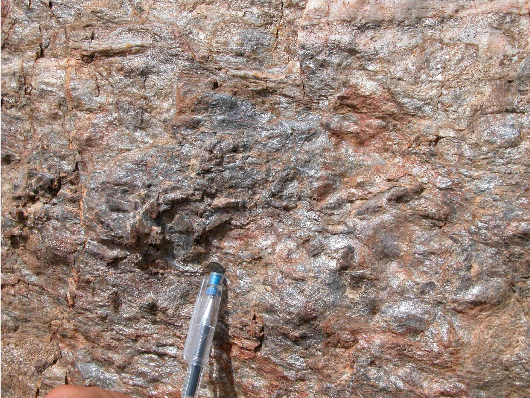
Stop 4.6- Road from Siniscola to Lodè; Mt. Bruncu Nieddu. Staurolite and garnet bearing micaschists. (Coord. N40° 36’ 0.6’’; E009° 34’ 56.6’’)
We stop few hundred meters north of the contact between orthogneisses and micaschists. At the entrance of a small forestal road, near the gate, we can observe cm-size staurolite and garnet porphyroclasts on the S2 foliation of the micaschists (Figure 4.15). The porphyroclasts of garnet, staurolite and plagioclase, grown between D1 and D2 deformation events, so that they are flattened, rotated and sometimes reduced in grain size during the D2 event (Figure 4.16)(Figure 4.17)(Figure 4.18)(Figure 4.19)(Figure 4.20) and (Figure 4.21). This is regarded as the first appearence of staurolite in northeastern Sardinia (Franceschelli et al., 1982, 1986, 1989a; Carmignani et al., 1982, 1994; Ricci et al., 2004 with references) marking the staurolite-in isograd. However, recent investigations revealed that staurolite + biotite appears nearly 10 km south of the classical isograd pointing to a more complex interplay between Barrovian metamorphism and defornation in northeastern Sardinia.
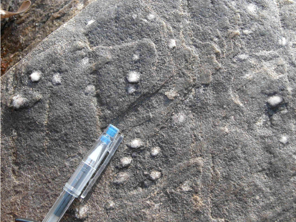
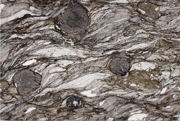
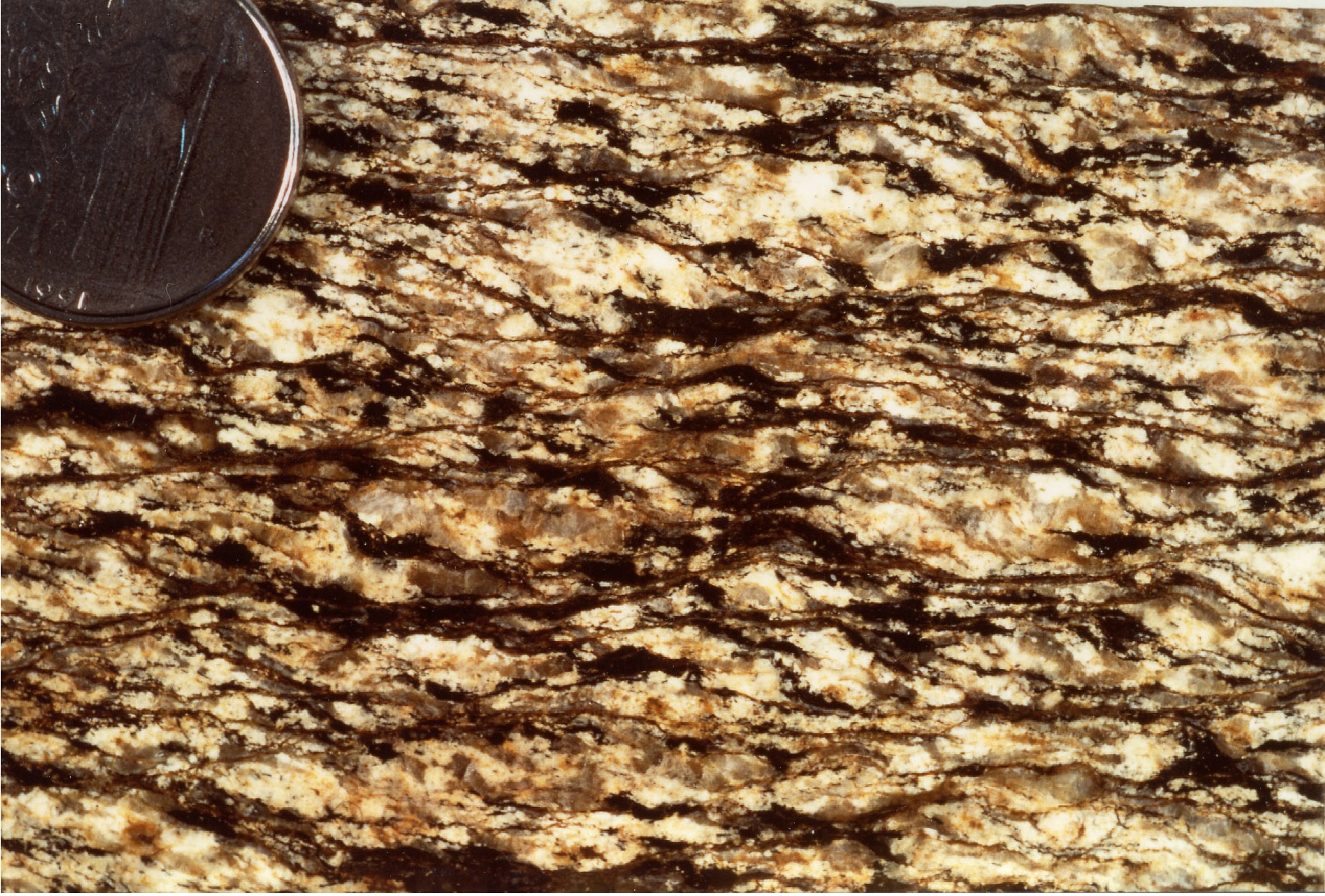
Figure 4.15. Cm-size staurolite porphyroclasts
Cm-size staurolite porphyroclasts developed on the S2 foliation in micaschsits at Stop 4.5.
Figure 4.16. Shear band cleavage in mylonites
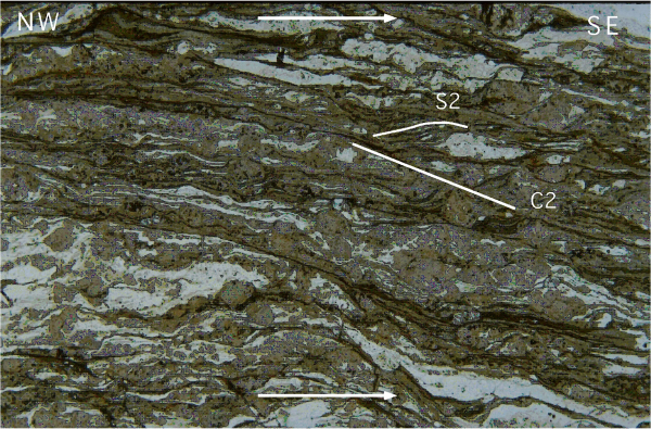
Shear band cleavage in mylonites from gneisses in the high-strain zone in the Posada Valley (top-to-the NW sense of shear, PPL; fov is nearly 7.7 mm) (Stop 4.5).

Figure 4.18. Thin section from micaschist
Thin section from micaschist of the St+Bt zone (Staurolite is the brown-yellowish mineral) with shear bands developed during D2 shearing (top-to-the NW sense of shear, PPL; fov 30 mm) (Stop 4.6).
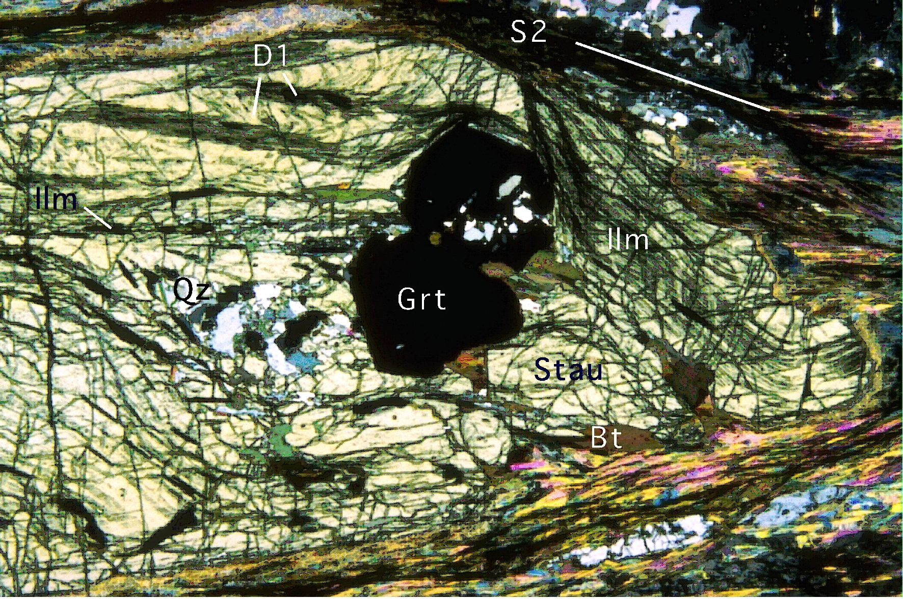
Figure 4.19. Photomicrograh showing stretched staurolite

Photomicrograh showing stretched staurolite (Stau) porphyroblast, grown post-D1 and pre-D2, including D1 related minerals (Grt, Ilm, Bt, Qtz) (PPL; fov is nearly 1.8 mm) (Stop 4.6).
Figure 4.20. Photomicrograh showing stretched staurolite porphyroblast
Photomicrograh showing stretched staurolite porphyroblast during D2 shearing (CPL; fov is nearly 2 mm) (Stop 4.6).
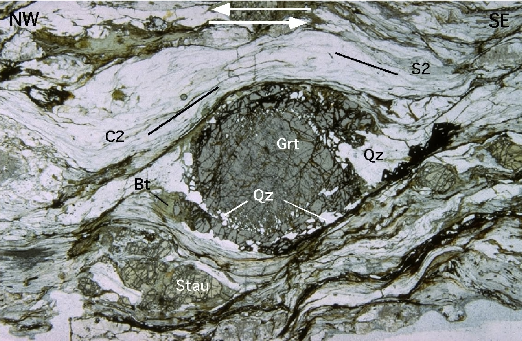
Figure 4.21. Syn-D2 rotated garnet (Grt) and relic staurolite

Syn-D2 rotated garnet (Grt) and relic staurolite (Stau): top-to-the NW sense of shear (micaschist from the St+Bt zone, PPL; fov is 4.5 mm) (Stop 4.6).
Staurolite porphyroclasts are often fractured and stretched along the S2 foliation with quartz, biotite, chlorite and white mica growing in the separated fragments.
The D1 fabric is transposed by D2 and only the S2 foliation that strikes W-E and WNW-ESE and moderately to strongly dips toward the S and the SW can be identified. It bears an oblique sub- horizontal stretching lineation (L2) marked by the alignment of chlorite, muscovite, quartz and biotite and by the stretched and fractured porphyroclasts of K-feldspar, kyanite, staurolite (Figure 4.21) and garnet. The rocks of the staurolite+biotite zone consist of staurolite, garnet and plagioclase porphyroblasts (up to 0.5 cm in size) often in a mylonitic matrix made up of phyllosilicates and quartz. Garnet is anhedral and rounded with quartz, biotite, and chlorite inclusions. Garnet shows a bell-shaped zoning from core to rim for Mn (Carosi & Palmeri, 2002). Towards the rim Mg and Fe gradually increase and Ca decreases. Staurolite occurs as elongated prisms with several phyllosilicates, quartz and graphite inclusions. Staurolite is chemically homogeneous and Fe-rich. Mg-content is up to 0.30 a.p.f.u. and XMg ratio ~ 0.17. XMg of biotite is 0.35. Muscovite is celadonite-poor (Mg+Fe up to 0.42).
Temperatures and pressures in the range of 570-625 °C and 0.7-1.0 GPa respectively have been reported by Franceschelli et al., (1989) and Di Vincenzo et al., (2004) for the thermal peak. In situ argon ages on muscovite along S2 foliation are mainly within 310-320 Ma (Di Vincenzo et al., 2004).
Moving northward for nearly 0.5 km along the forestal road we reach kyanite+biotite isograd.
The kyanite+biotite isograd is marked by the first appearance of kyanite crystals. The rocks consist of porphyroblasts of staurolite, kyanite, and plagioclase enveloped in a mylonitic matrix of muscovite, biotite, chlorite and ilmenite. Garnet often occurs as euhedral clear or cloudy inclusion in staurolite, plagioclase and rarely in kyanite porphyroblasts. The clear garnet contains calc- silicate micro-inclusions of idiomorphic anorthite, epidote and margarite. Garnet from the matrix or as a cloudy inclusion have a similar composition, with a slight increase in spessartine content from core to rim, and a concomitant decrease in the other garnet components. Plagioclase included in garnet is extremely calcic (An= 99-67) while the one enclosed in cloudy garnet has a compositional range of An=22-59. XMg of biotite ranges from 0.8 to 1.2 and TiO2 is up to 2.3%. Muscovite is Na- and celadonite poor (Ricci et al., 2004).
Temperature up to 595 °C and pressures up to 0.67 GPa have been reported by Franceschelli et al. (1989) and pressures > 0.9 GPa by Carosi & Palmeri (2002) for the metamorphic peak.
The micaschists are intruded by an undeformed Permo-Triassic dyke of camptonite (K-Ar age: 228?3 Ma form Baldelli et al., 1987) with mineralogical and textural feature of a lamprophyre. It is porphyritic with biotite and amphibole euhedral phenocristals.
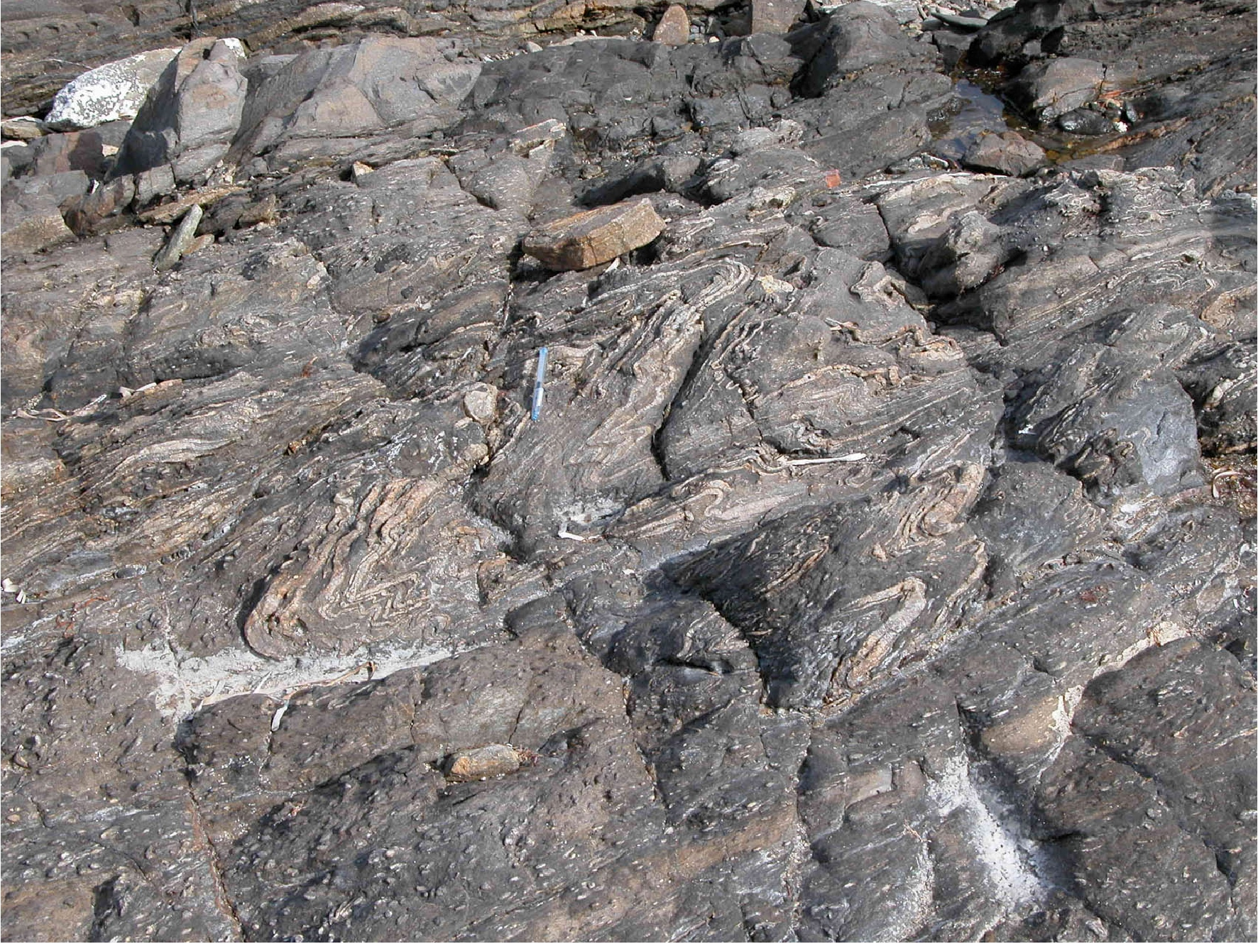
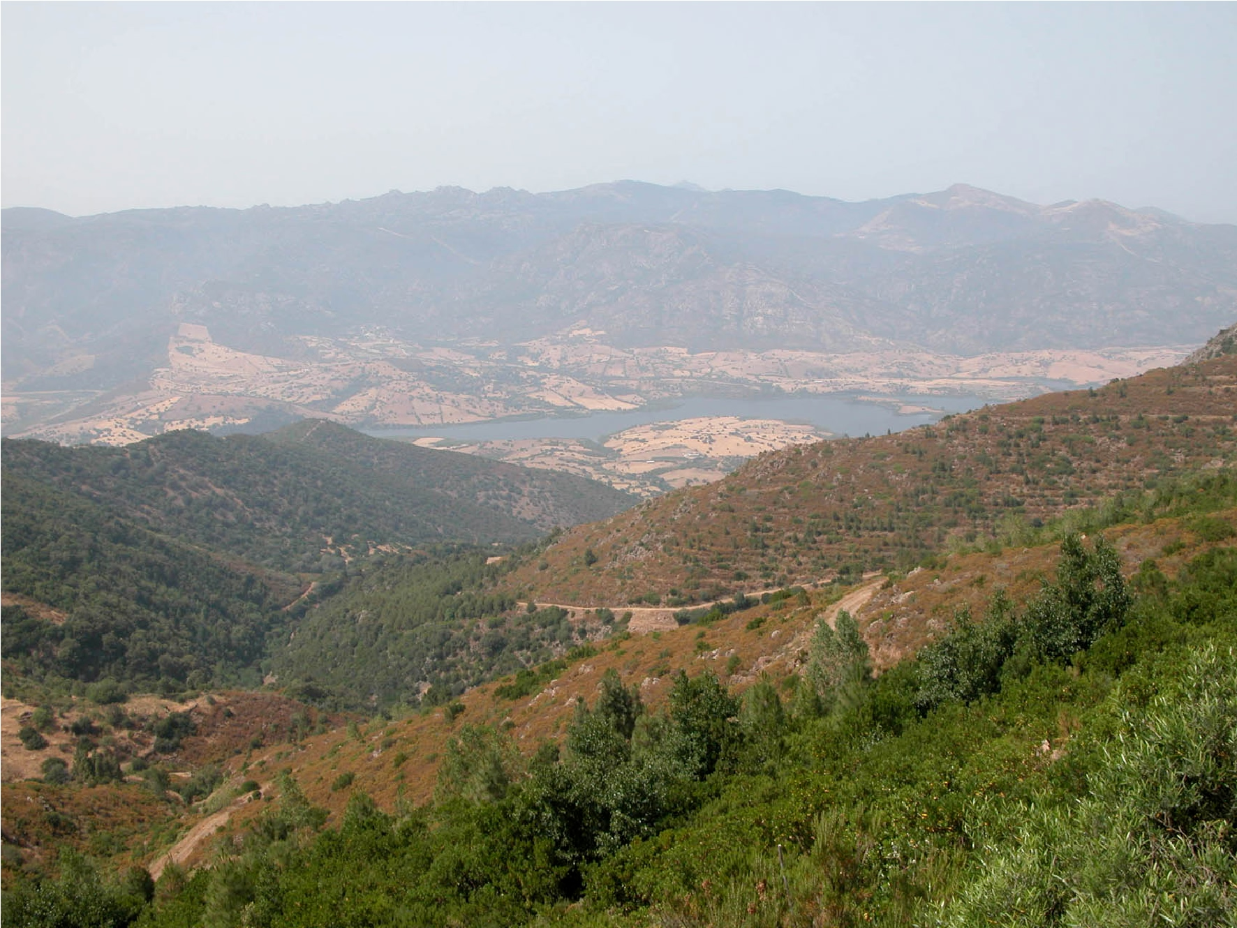
At the bottom of the Rio Posada valley (Figure 4.22) there is the sillimanite-muscovite isograd roughly coinciding with the first appearance of migmatitic rocks, characterized by quartz-plagioclase-muscovite bearing leucosomes. Franceschelli et al., (1989) estimated a temperature of 605 °C and pressure of 0.4 GPa for this zone.
Figure 4.22. Overview from south to north of the Posada Valley from Stop 4.5
In front of us there are mylonites form staurolite and kyanite bearing micaschists and paragneisses and going down to the Posada Valley we encounter granites and migmatites which made up the far mountains in the far field.
Going into the migmatites we enter in sillimanite+muscovite and in sillimanite+k-feldspar zone. The oldest structure observed in migmatites is a gneissose layering, pre-dating the most pervasive (S2) foliation. Mesosomes are medium-grained, with a fabric defined by the alignment of biotite parallel to S2 schistosity. Mesosomes consist of quartz, plagioclase, biotite, garnet, fibrolite, minor kyanite, muscovite and K-feldspar. Kyanite occurs sporadically as relic minerals. Retrograde white mica occurs in both the mesosome and the leucosome on sillimanite and K-feldspar. Leucosomes are coarse-grained, poorly-foliated rocks, tonalitic to granitic and rarely trondjemitic in composition.
Temperatures up to 750 °C under which anatexis processes developed and pressures at 0.6-0.8 GPa have been reported for migmatites (Palmeri, 1992; Cruciani et al., 2001). According to Giacomini et al., (2005) migmatization started in the kyanite stability field at about 750-800°C and pressures above 1.0 GPa.
Argon ages on muscovites from both migmatitic orthogneiss and metasedimentary stromatic migmatite from Punta de li Tulchi yielded a strongly comparable and restrict age-variation from ~300 Ma to ~320 Ma (Di Vincenzo et al., 2004).
It is worth to note that in the classical view of the Barrovian metamorphism in northern Sardinia the appearing of sillimanite has been put in a prograde metamorphic sequence after kyanite (Franceschelli et al., 1982, 1986, 1989 and Ricci et al., 2004 with references therein). It is certainly true for temperature but not for pressure that shows decreasing values of at least 0.3-0.4 GPa passing from the kyanite to the sillimanite+muscovite zone (see Carosi & Palmeri, 2002 and Ricci et al., 2004). Moreover, in this area, in SW Gallura and Asinara Island the sillimanite starts to grow along the S2 foliation whereas porphyroblastic staurolite and kyanite grew before S2 foliation. We therefore suggest that the prograde Barrovian metamorphism reached the kyanite zone and from broadly during D2 medium pressure metamorphic rocks and migmatites were decompressed (isothermal decompression in the migmatites according to Carosi et al., 2004 and Giacomini et al., 2005) and subsequently reached the sillimanite stability field.
So instead of a classical entire Barrovian sequence we consider the basement of northern Sardinia affected by a more complex metamorphic evolution characterized by an earlier prograde metamorphism of broadly Barrovian type (or even with higher P/T ratios) acquired during the underthrusting of continental crust reaching the higher pressures in the kyanite stability field followed by a nearly isothermal decompression from D1 to D2 reaching the sillimanite stability field, followed in some places (e.g. SW Gallura and Asinara island) by a further HT-LP metamorphic event.
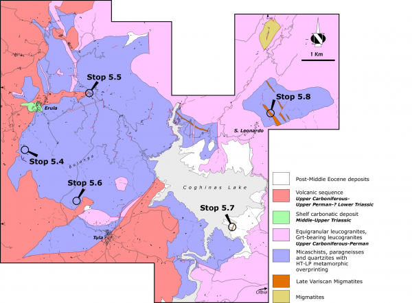
Itinerary 5: The Migmatitic Complex in SW Gallura region and the HT-LP metamorphic overprint in Anglona
The Migmatitic Complex in SW Gallura
Stop 5.1- The Migmatitic Complex is characterized by a deep alteration that locally erases completely the migmatitic texture. Along the main street from Viddalba to Aggius we can observe the dominant lithotype in a little quarry on the right where there are metatessites with irregular and millimetric-thick leucosomes deeply fractured and locally folded by F2 and F3 fold systems.
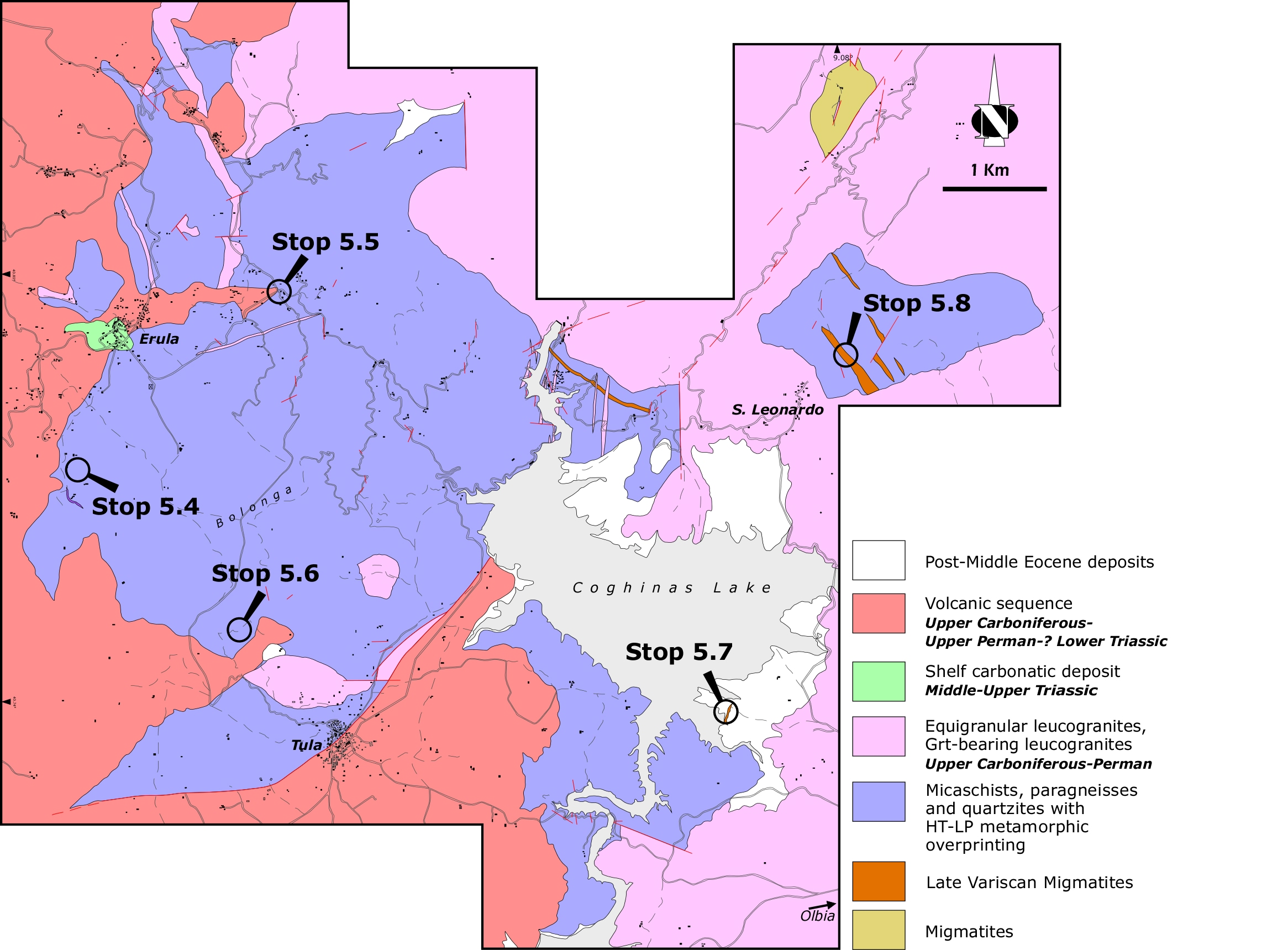
Figure 5.1. Geological map of the Migmatitic Complex
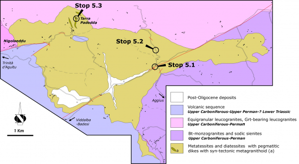
Geological map of the Migmatitic Complex in SW Gallura and indication of the stops.
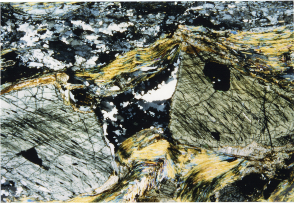
Stop 5.2- Turning on the left we can observe well developed pegmatitic veins generally parallel to the S2 foliation associated with small leucosome (Ashworth, 1985) with millimetric melanosomes (Figure 5.2A). Rare migmatitic gneisses have been recognized.
Figure 5.2. The Migmatitic Complex in SW Gallura region (Stop 5.1)
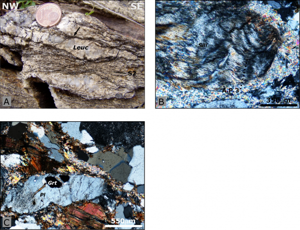
A. Thin restitic layers around the major leucosome (arrow); B. Fibrolite folded by D3 deformation phase (CPL); C. Idioblastic garnets in plagioclase (CPL).
The leucosomes show a trondjemitic composition with quartz and plagioclase. This implies that these migmatites represent the first melting event in Sardinia that could be related both to metamorphic differentiation and to partial melting (Del Moro et al., 1991; Oggiano & Di Pisa, 1992; Di Vincenzo et al., 2004). Fibrolite seems to be folded by F3 folds (Figure 5.2B) whereas rare relics of Pl, Grt (inside Pl) testify the earlier lower metamorphic conditions (Figure 5.2C).
Coming back to Viddalba we turn on the first street on the right toward Nigolaeddu village.
The HT-LP metamorphic overprinting in the Anglona region
The Coghinas Lake Complex (CLC) (Anglona region) represents the southernmost metamorphic complex in this sector of axial zone. This complex has been interested by an important HT-LP metamorphic overprinting that erases, almost always, its sedimentary nature.
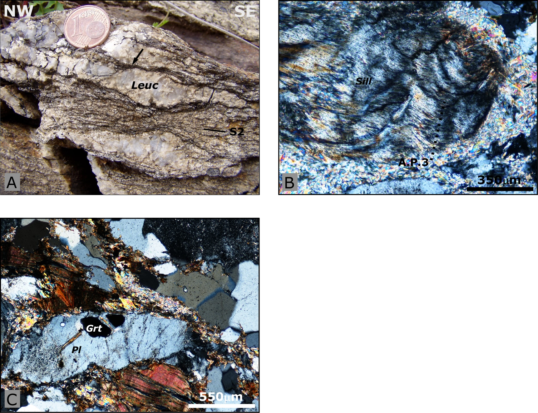
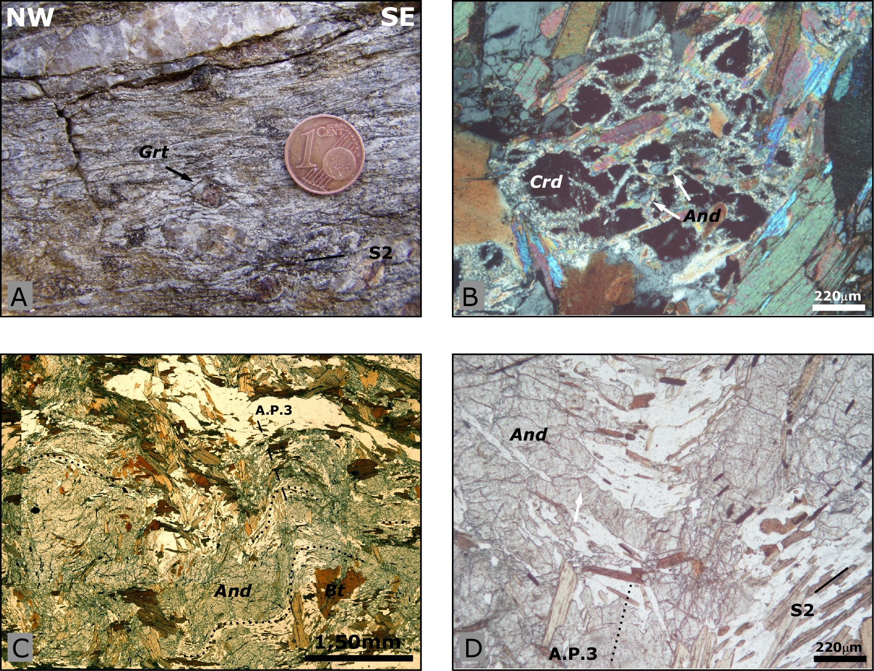
Stop 5.4- Near Erula village stadium relics of S1 foliation can be observed inside centimetric D2 microlithons developed in more quartzitic levels.
Millimetric garnet crystals, enveloped by S2 foliation, and characterized by a reaction rim of biotite testify the previous Barrovian metamorphism (Figure 5.4A).
Figure 5.4. The HT-LP metamorphic overprinting in the Anglona region (Stop 5.5)

A. Garnet (Grt) with well developed reaction rim of biotite; B. Cordierite (Crd) developed on andalusite crystal (CPL); C. Andalusite sin/post-D3 (PPL); D. Photomicrograph of post-D3 andalusite crystal (And) (A.P.3: axial plane of third deformational phase) (plane polarized light).
Stop 5.5- On the Sa Mela locality we can observe the typical lithotype outcropping in this metamorphic complex. The main foliation is highlighted by millimetric alternation of biotite and quartz-feldspatic layers and by the presence of injections of quartz generally parallel to the main foliation (often interested by F2 and F3 folds).
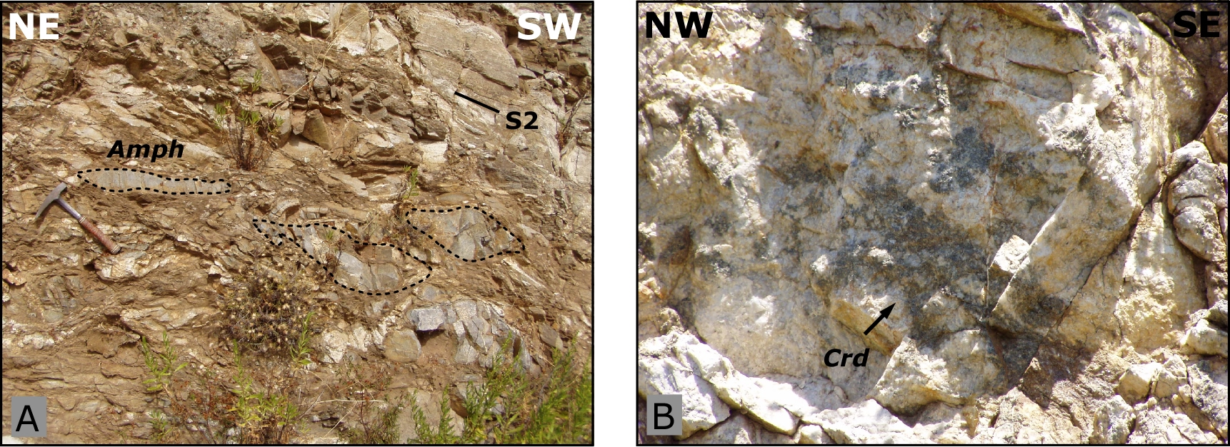
Figure 5.5. The migmatites in Anglona region (Stop 5.8)

A. Amphibolitic bodies (Amph) in the S. Leonardo area; B. Cordierite (Crd) nodules.
The main mineral association is represented by biotite, plagioclase, quartz, muscovite, sillimanite, garnet, chlorite, rutile and zoisite. Andalusite and cordierite blastesis has been developed locally. Their textural analyses testify that deformation developed during high-grade metamorphic conditions.
Centimetric crystals of andalusite growth statically on limbs of F3 folds. Cordierite is probably coeval with andalusite (Figure 5.4B) and it develops from reactions St+Qtz=Crd+Als+H2O (Ricci, 1992) after or during late stages of D3 deformation phase (Figure 5.4C and D) 5.4C and D).
Stop 5.6- Coming back to Erula, we turn on the left on the road to Tula across the Bolonga plateau. Near the Mt. Nieddu we can see mm-thick quartz-feldspatic layer folded by F2 folds and interested by top-to-the SE shear zones developed during the later stage of F2 fold development. An analogous structure has been recognized also in the southeastern side of Mt. Nieddu and in the northern portion of the metamorphic complex.
Arrived in Tula we take the Olbia-Alghero road to observe the migmatites developed at the expense of the HT-LP micaschist in the eastern side of the Coghinas Lake.
Stop 5.7- On the SE shore of the Lake we can observe F2-F3 interference folds system, relics of S1 foliation and small gneiss body. Migmatites, clearly developed after the HT-LP metamorphic overprinting, show generally agmatitic and diatessitic texture.
Stop 5.8- In the S. Leonardo region there are small amphibolitic bodies (Figure 4.5A) and well-developed metatessites with decimetric leucosomes and nodules of cordierite, partly preserved (Figure 4.5B). These migmatites develop from the HT-LP micaschists.