Microstructures of rodingites
Three types of rodingites have been distinguished on the basis of their main mineral constituents, epidote, garnet, clinopyroxene, chlorite and vesuvianite.
Epidote-bearing rodingites consist of whitish layers of epidote and greenish layers of clinopyroxene, locally containing mm-sized greenish chlorite lenses. Up to cm-sized grey to pale green clinopyroxene porphyroclasts occur, whereas vesuvianite is absent.
Garnet-chlorite-clinopyroxene-bearing rodingites are pale pink to red or rarely green rocks, whose foliation is defined by up to 1 or 2 cm-thick chlorite- and garnet-rich layers. Whitish grey aggregates of clinopyroxene and rare epidote and garnet are parallel to the foliation. Clinopyroxene porphyroclasts show a grey violet colour and are up to 5 cm-sized (Fig. 7). Bright green aggregates made of Cr-garnet and spinel may occur. Vesuvianite + epidote never exceed 10% in volume.
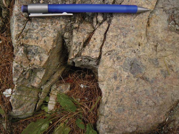
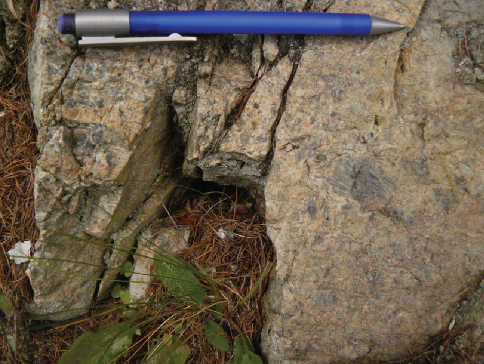
Figure 7. Relict magmatic texture in rodingites
Cm-sized clinopyroxene porphyroclasts in a rodingite boudin south of Crépin, close to the contact with metagabbros. The composite S1/S2 foliation wraps the porphyroclasts.
Vesuvianite-bearing rodingites are characterised by a micro-crystalline green matrix consisting of chlorite, garnet, and clinopyroxene in variable proportions. Locally there are cm-sized clinopyroxene porphyroclasts aligned parallel to the composite S1/S2 foliation. In places garnet- and calcite-bearing veins are positioned at a high angle to boudin elongation.
Epidote-bearing rodingites
The main mineral phases are: epidote (20 - 70%), clinopyroxene (15 - 50%), Mg-chlorite (10 - 20%), garnet (≤ 20%), tremolite (≤ 5%).
S1 foliation is defined by SPO (Shape Preferred Orientation) of epidoteI and by LPO (Lattice Preferred Orientation) of Mg-chloriteI and clinopyroxeneI. EpidoteI (locally Fe-epidote) forms euhedral zoned, coarse-grained, and twinned crystals. GarnetI is enclosed in, and shows rational rims with epidoteI (Fig. 8a). ClinopyroxeneI and minor epidoteI form aggregates parallel to S1. The margins of clinopyroxeneI show inclusion trails marked by opaque minerals in continuity with, and sub-parallel to the external S1.
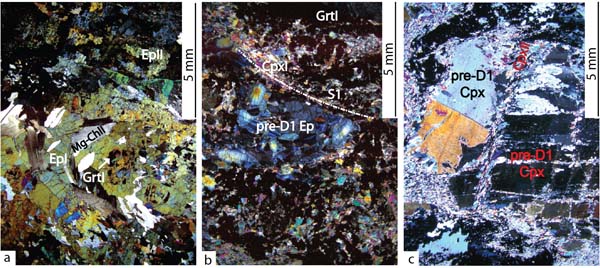
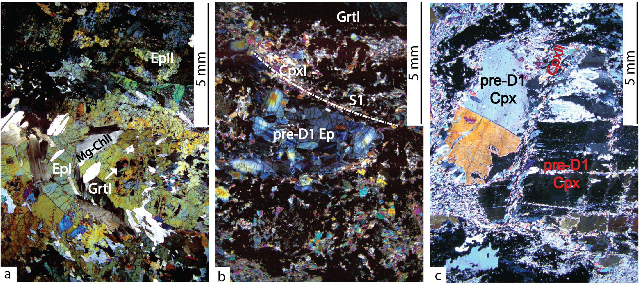
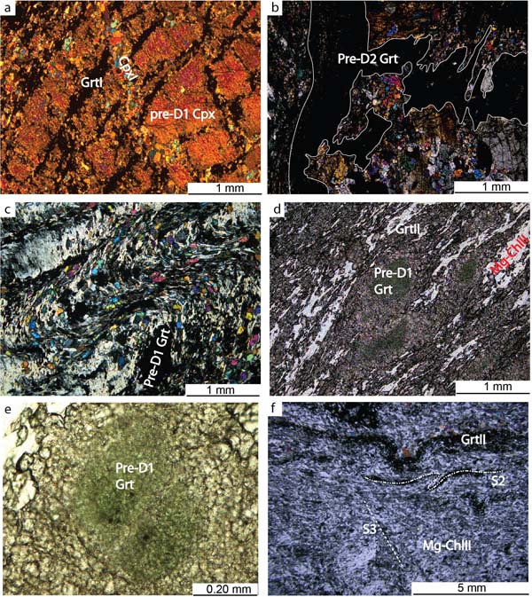
Figure 8. Pre-Alpine and Alpine microstructures in Epidote-bearing rodingites.
Mineral compositions related to microstructural sites are shown in Figs. 12, 13, and 14 and in Tables 1 and 2. a) D2 folding deforms Mg-chloriteI and epidoteI that dynamically recrystallise. GarnetI is enclosed in epidoteI. b) S1 foliation, marked by Mg-chloriteI, garnetI, and clinopyroxeneI, wraps around an epidote aggregate. Crossed polars. c) clinopyroxeneII in the fractures and along cleavages of a Pre-D1 clinopyroxene porphyroclast. Crystal-plastic shear deformation is accommodated in clinopyroxeneII, garnetII, and minor tremoliteI multi-granular aggregate filling fractures. Crossed polars.
S2 is defined by SPO and/or LPO of clinopyroxeneII, garnetII, Mg-chloriteII, epidoteII, and rare tremoliteI. EpidoteII, often forming polygonal aggregates, is also enclosed in Mg-chloriteII. Veins consisting of pre-D2 garnet and minor pre-D2 epidote are folded by D2 and may be locally transposed into the folded S1. Mg-chloriteI forms porphyroclasts within S2. TitaniteI forms thin trails parallel to S2 and rarely occurs in clinopyroxeneII layers.
Where S2 is more developed epidoteII (zoisite and clinozoisite) is in fine-grained crystals forming trails parallel to S2 with clinopyroxeneII and garnetII.
Up to 1 cm-sized kinked pre-D1 clinopyroxene porphyroclasts show SPO either parallel to S1 or S2. These porphyroclasts locally have opaque mineral exsolutions and are overgrown by rims of clinopyroxeneI/II at the edges and along fractures. These rims do not contain opaque minerals, are optically continuous with the pre-D1 clinopyroxene porphyroclasts, and form spikes parallel to S2 foliation, suggesting that pre-D1 clinopyroxene porphyroclasts partially recrystallised during D2. The cores of pre-D1 clinopyroxene porphyroclasts are locally replaced by Mg-chlorite along cleavages.
Pre-D1 clinopyroxene porphyroclasts enclose pre-D1 epidote crystals that also form aggregates wrapped by S1; the core of these epidote crystals is Fe-rich, as suggested by high birefringence colours (Fig. 8b); epidote strain shadows are filled by Mg-chloriteI or II. In more foliated rocks pre-D1 clinopyroxene porphyroclasts are fractured within S2, with mineral filling of clinopyroxeneII, garnetII, and rare tremoliteI (Fig. 8c). The high angle with S2 and SPO of clinopyroxeneII filling these fractures, suggest that they developed during D2. These fractures are intersected by other fractures at a high angle with S2 that are filled by coarse-grained euhedral epidoteII crystals. Fractures cutting S2 are filled by epidoteIII, which is zoisite and clinozoisite where fractures intersect epidoteI or epidoteII and Fe-epidote where fractures intersect garnet, clinopyroxeneII, and Mg-chloriteII aggregates.
Rare post-D2 tremoliteII forms randomly oriented crystals overgrowing garnet and epidote layers. Rocks containing tremolite are the poorest in epidote.
Taking into account the described microstructural features, the following mineral growth - deformation relationships can be inferred in epidote-bearing rodingites:
- Pre-D1 mineral relics: Cpx porphyroclasts, ± Ep (Fe-Ep);
- Syn-D1: EpI, CpxI, MgChlI, GrtI;
- Syn-D2: EpII, CpxII, MgChlII, TtnI, ± GrtII, ± TrI;
- Post-D2: ± EpIII, ± TrII.
Garnet-chlorite-clinopyroxene-bearing rodingites
Garnet-chlorite-clinopyroxene-bearing rodingites consist of: garnet (20 - 70%), Mg-chlorite (20 - 55%), clinopyroxene (≤ 50%), opaque minerals (≤ 5%), vesuvianite (≤ 10%).
These rocks are heterogeneously deformed and the dominant fabric can be the S1 foliation, the crenulated S1 with the newly forming variably differentiated S2 crenulation cleavage, or the fully decrenulated continuous S2 foliation.
S1 is marked by garnetI, Mg-chloriteI, clinopyroxeneI, and rare prismatic vesuvianiteI. In samples where S1 is pervasive, rare vesuvianite porphyroblasts, postdating S1, enclose fine-grained garnetI. Up to 1 cm-sized pre-D1 clinopyroxene porphyroclasts have fractures filled mainly by fine-grained garnetI and/or clinopyroxeneI showing complex relations: fractures filled by clinopyroxeneI ± epidoteI are intersected and displaced by fractures filled by garnetI ± Mg-chloriteI or garnetI fractures are intersected by clinopyroxeneI fractures (Fig. 9a). Where fractures form a high angle with S1, the mineral filling may be interpreted as syn-D1; in other cases the relative chronology of mineral growth filling with respect to the deformation stages is not clear. Pre-D1 clinopyroxene porphyroclasts are rimmed by garnetI, which also grew in continuity along the S1 films. GarnetI and minor clinopyroxeneI + Mg-chloriteI also form aggregates, locally parallel to S1, which could be pseudomorphs after pre-D1 clinopyroxene porphyroclasts. The aggregates are affected by ductile boudinage with Mg-chloriteI in the necks. Two sets of conjugate micro-shear zones marked by Mg-chloriteII and rare vesuvianiteII affect S1 and kink Mg-chloriteI.
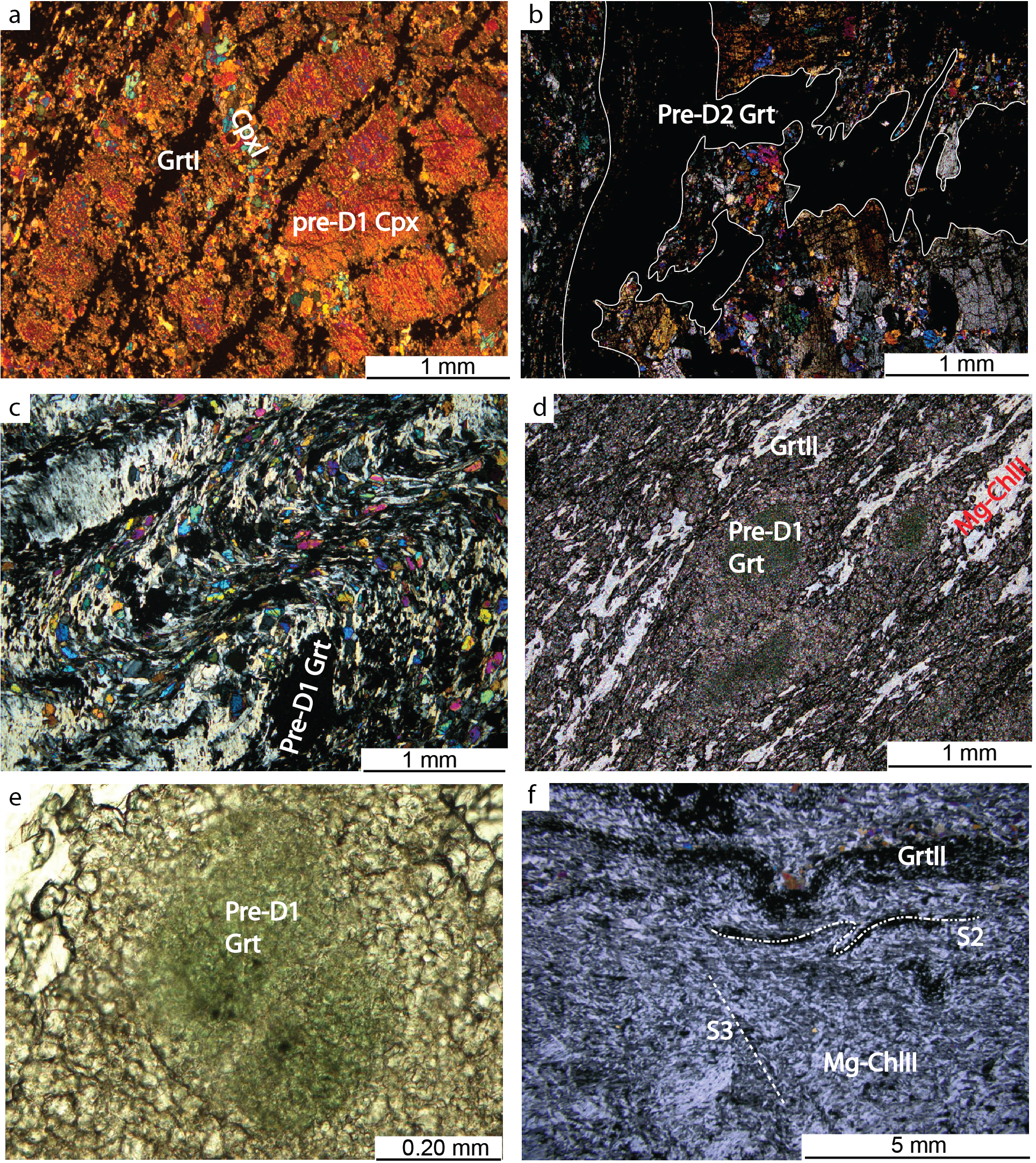
Figure 9. Pre-Alpine and Alpine microstructures in Garnet-chlorite-clinopyroxene-bearing rodingites.
Mineral compositions related to microstructural sites are shown in Figs. 13 and 14 and in Tables 1 and 2. a) Pre-D1 clinopyroxene porphyroclast transected by garnetI-filled fractures and by clinopyroxeneI-filled fractures. Crossed polars. b) Microfolding and transposition along S2 of a pre-D2 garnet-vein; white line highlights vein margins; crossed polars. c) S1 and the differentiated S2 marked by garnetI/II, Mg-chloriteI/II, and clinopyroxeneI/II. An uvarovite-rich pre-D1 garnet porphyroclast is preserved within the crenulated S1; crossed polars. d) Uvarovite-rich garnet wrapped by S2 foliation, marked by garnetII, chloriteII, and clinopyroxeneII; plane polarised light. e) Uvarovite-rich garnet porphyroclast with Cr-spinel (dark spots) preserved in the core: plane polarised light. f) Folded S2 defined mainly by Mg-chloriteII and garnetII-rich aggregates. Mg-chloriteIII concentrates along the incipient S3 differentiated cleavage; crossed polars.
Pre-D2 garnet, clinopyroxene, and minor Mg-chlorite fill pre-D2 veins, which are parallel to or folded and at a high angle with S2 (Fig. 9b).
Where S2 is only incipient, it is a spaced crenulation foliation with folded S1 relicts within the S2 films. S1 and S2 are marked by SPO of clinopyroxeneI and II and garnetI and II and SPO and LPO of Mg-chloriteI and II respectively (Fig. 9c). Some clinopyroxeneII grains show an internal foliation defined by opaque mineral that is slightly folded and in continuity with the external S2, suggesting that these grains are syn-kinematic to S2.
Locally S1- or S2-parallel garnetI/II aggregates wrap pre-D1 garnet porphyroclasts, which have a green Cr-rich core and colourless rims recrystallised during D1 or D2 (Fig. 9d). The Cr-rich core may contain fine-grained Cr-rich spinel (Fig. 9e). When the pre-D1 garnet aggregates are parallel to S1 they are dismembered and folded by D2.
Well-developed S2 is marked by LPO and SPO of Mg-chloriteII, garnetII, clinopyroxeneII, and rare opaque minerals; clinopyroxeneII forms layers parallel to S2.
Pre-D1 clinopyroxene porphyroclasts may also show SPO parallel to S2. In this case garnetII crystallised in fractures at a low angle with clinopyroxene porphyroclast cleavages and in continuity with S2. In wider fractures Mg-chloriteII forms crystals oriented at about 45° with respect to the fracture wall, suggesting wall-parallel shearing.
In D2 microfolds, Mg-chloriteI forms aggregates of recrystallised decussate grains or is internally deformed. Mg-chlorite porphyroclasts are overgrown by garnetII along rims and cleavages and may be related to D1 or pre-D1 stages. Euhedral opaque minerals overgrow S2.
Mg-chloriteIII marks the incipient S3 crenulation cleavage of locally asymmetric micro-folds (Fig. 9f). Rare Mg-chloriteIII intersects S2 films at low angle.
The microstructural features described above indicate the following mineral growth - deformation sequence:
- Pre-D1 mineral phases: uvarovite-rich Grt, Cpx porphyroclasts, ± Cr-rich Spl, ± Mg-Chl
- Syn-D1 or pre-D2: Mg-ChlI, GrtI, ± CpxI, ± EpI, ± VesI, ± opaque minerals
- Syn-D2 or post-D1: Mg-ChlII, GrtII, CpxII, ± VesII, ± opaque minerals
- Syn-D3: Mg-ChlIII.
Vesuvianite-bearing rodingites
Vesuvianite-bearing rodingites consist of: vesuvianite (25 - 55%); Mg-chlorite (10 - 55%); garnet (10 - 30%); clinopyroxene (5 - 20%).
S2 foliation is marked by fine-grained garnetII, SPO of zoned vesuvianiteII and clinopyroxeneII, and LPO and SPO of Mg-chloriteII (Fig. 10a). S2 is also marked by layers of different grain size and modal proportions of vesuvianiteII, clinopyroxeneII, or garnetII crystals with rare titaniteI films and minor epidoteI.
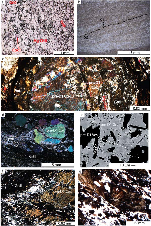
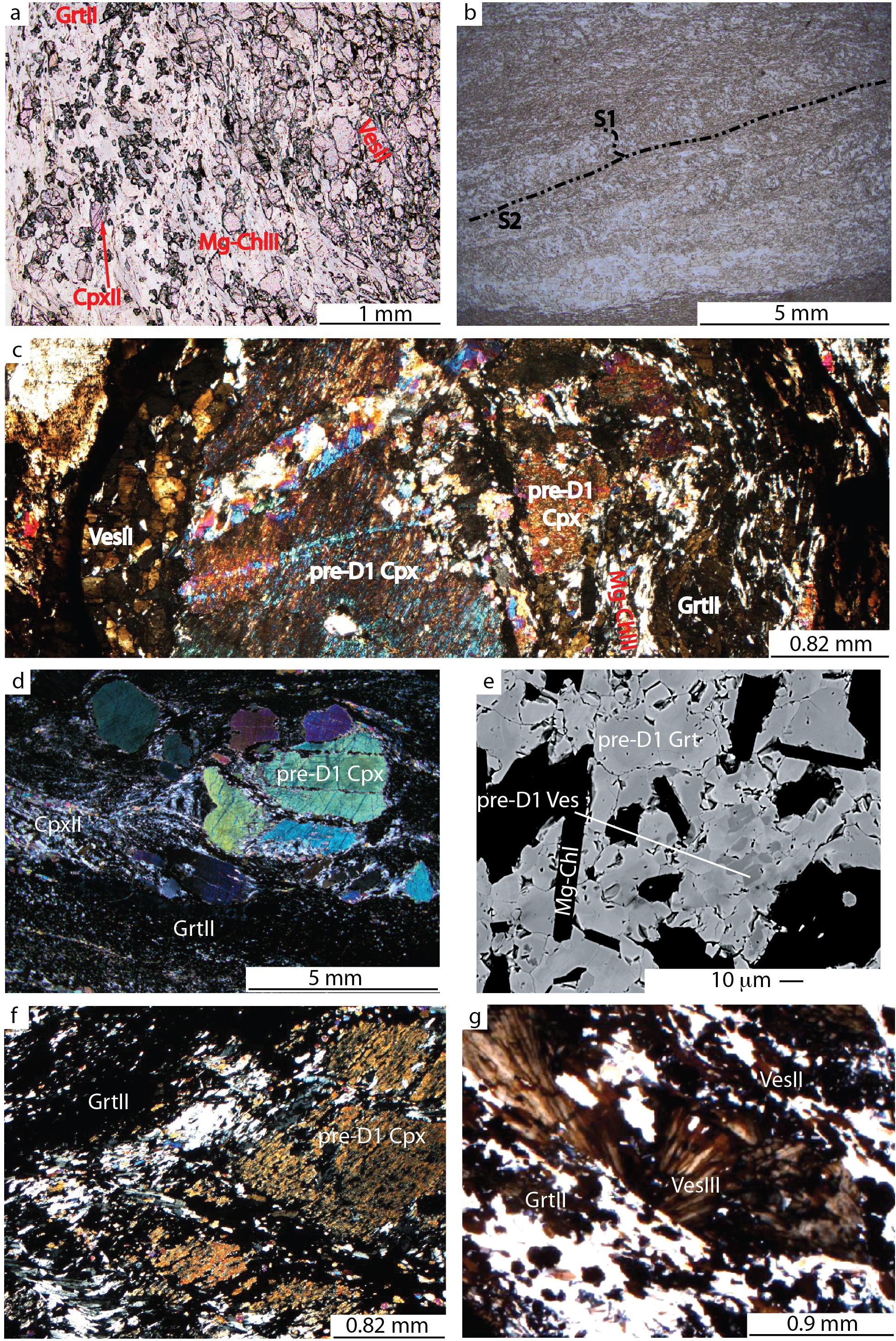
Figure 10. Pre-Alpine and Alpine microstructures in vesuvianite-bearing rodingites.
Mineral compositions related to microstructural sites are shown in Figs. 13, 14, and 15 and in Tables 1 and 2. a) VesuvianiteII, garnetII, Mg-chloriteII, and minor clinopyroxeneII marking S2; plane polarised light. b) S1 geometry preserved within S2 lithons; stages of progressive enrichment of vesuvianiteII, Mg-chloriteII, and garnetII are evident by variable thickness of S2 films; plane polarised light. c) Clinopyroxene porphyroclast wrapped by and aligned along the S2 foliation, marked by vesuvianiteII, garnetII, Mg-chloriteII, and clinopyroxeneII. New clinopyroxene crystallised at the rim and in fractures of the porphyroclast. Vesuvianite marking S2 is zoned (see also Fig. 15); crossed polars. d) Fractured pre-D1 clinopyroxene porphyroclast wrapped by S2; Mg-chloriteII crystallised mainly in the strain shadows and Mg-chloriteII and garnetII filled the fractures. New clinopyroxeneII is concentrated in the layer at the tip of the strain shadows; crossed polars. e) Pre-D1 vesuvianite enclosed in the core of pre-D1 garnet; backscattered SEM image. f) Pre-D1 clinopyroxene porphyroclast replaced by garnetII and minor clinopyroxeneII that grows along cleavages and fractures; crossed polars. g) VesuvianiteIII overgrowing the S2 foliation marked by vesuvianiteII, Mg-chloriteII, and garnetII; crossed polars.
Rare pre-D2 clinopyroxene and Mg-chlorite are oblique to S2 and locally form aggregates wrapped by S2. Mg-chloriteI forms decussate aggregates of re-crystallised grains within S2 films, evidencing an S1 relict foliation. S1 is sporadically preserved between S2 films as a crenulated foliation (Fig. 10b) mainly marked by Mg-chloriteI, vesuvianiteI, and garnetI.
Mm-sized pre-D1 clinopyroxene porphyroclasts are wrapped by S2 and may show SPO parallel to S2 (Figs. 10c, d). Minor epidote may be present as subhedral crystals at the pre-D1 clinopyroxene porphyroclast rims. Very fine-grained opaque minerals are exsolved from the pre-D1 clinopyroxene porphyroclast crystal lattice whereas the porphyroclast rims (clinopyroxeneII) do not contain opaque minerals. Strain shadows are filled by vesuvianiteII and Mg-chloriteII. Cr-rich pre-D1 garnet porphyroclasts are wrapped by S2 foliation and contain euhedral to subhedral crystals of pre-D1 vesuvianite; therefore pre-D1 garnet and vesuvianite are in contact by rational margins, suggesting textural equilibrium (Fig. 10e).
Garnet and clinopyroxene also grew along rims and fractures of clinopyroxene porphyroclasts (Fig. 10f). In some samples fractures occur in two sets, located at a low and high angle with respect to S2; fractures in pre-D1 clinopyroxene porphyroclasts at a high angle with S2 can be interpreted as syn-D2. ClinopyroxeneII-bearing fractures cut garnetII ± Mg-chloriteII and vesuvianiteII filled fractures.
VesuvianiteIII forms crystals or garbens that overgrow S2 films (Fig. 10g). Mg-chloriteIII fills veins intersecting S2. Rare Mg-chloriteIII marks an incipient S3 axial plane foliation or intersects S2 at a low angle.
Summarising, the following mineral growth-deformation relationships can be inferred:
- Pre-D1 or pre-D2 minerals: Cpx porphyroclasts, ± pre-D1 Grt, ± pre-D1 Ves
- syn-D1 assemblage: Mg-ChlI, ± GrtI, ± VesI
- syn-D2 assemblage: VesII, Mg-ChlII, CpxII, GrtII, ± TtnI, ± EpI
- syn-D3 minerals: Mg-ChlIII, VesIII.
Rodingite/serpentinite reaction rims:
The reaction rim of rodingites consists of clinopyroxene and chlorite bearing schists.
S1 foliation is variably folded and mostly obliterated by the S2 crenulation cleavage. S1 is marked by a layering characterised by SPO and LPO of clinopyroxeneI and chloriteI (Fig. 11a); minor opaque minerals may be scattered along S1-parallel layers. ClinopyroxeneI may define layers characterised by a remarkably variable grain size. Locally disharmonic folding of chloriteI- and clinopyroxeneI-layers occurs. S2 crenulation cleavage is marked by LPO and SPO of clinopyroxeneII, chloriteII, and amphiboleI (Fig. 11b). ChloriteIII forms rare radial aggregates and randomly oriented amphiboleII overgrew S1 and S2 or mimetically replaced clinopyroxeneII.
The above described microstructures suggest the following mineral growth-deformation relationships for the reaction rims:
- D1: ChlI, CpxI, opaque minerals
- D2: ChlII, CpxII, AmpI
- Post-D2: AmpII, ChlIII
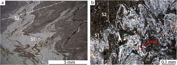
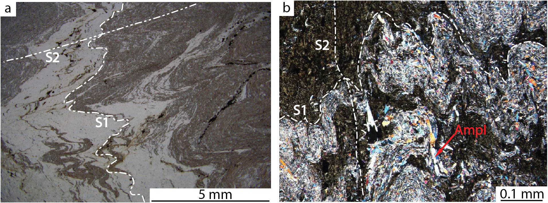
Figure 11. Alpine microstructures in rodingite reaction rims.
a) S1 is marked by a mineral layering of alternating clinopyroxeneI and chloriteI, and is crenulated during D2 deformation; plane polarised light. b) The S1 foliation is crenulated during D2. ClinopyroxeneI and chloriteI mark S1; the incipient differentiation of the S2 crenulation foliation is highlighted by amphiboleI crystals aligned with S2; crossed polars.