1. Introduction
Veins and strain fringes are two types of dilation structures in which new crystals have grown (Durney and Ramsay, 1973; Ramsay and Huber, 1983; Passchier and Trouw, 1996; Bons, 2000). Veins are elongate dilation sites which normally form due to inital fracturing of the host-rock whereas strain fringes grow next to rigid objects at the site of the extensional instantaneous stretching axes of flow (ISA). A fringe structure consists of two fringes and a rigid core-object. Syndeformational crystal textures in veins and strain fringes are of great importance for structural analysis since they are thought to record part of the progressive deformation history of the host-rock (e.g. Durney and Ramsay, 1973; Ramsay and Huber, 1983; Ellis, 1986; Passchier and Trouw, 1996). Grain boundaries of crystals with high length to width ratios (fibres) are thought to follow the opening trajectory of veins and strain fringes. However, there are a number of uncertanties in fibre analysis since not all fibres grow in the direction of vein or fringe opening (fig. 1, Cox and Etheridge, 1983; Fisher and Brantley, 1992; Bons and Jessell, 1997; Koehn and Passchier, 2000). Recently numerical experiments have been carried out to improve our understanding of crystal textures in dilation sites (Koehn et al., 2000, 2001; Bons, 2001; Hilgers et al., 2001) based on a crystal growth hypothesis put forward by Urai et al. (1991). In this paper we show some results of these studies in the form of movies in order to illustrate the progressive development of crystal textures in veins and strain fringes.
|
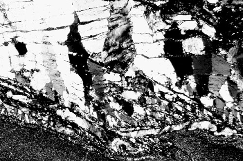
Figure 1. Crystal textures in a calcite, quartz and mica vein from the Orobic Alps (northern Italy). Crystals grew blocky to elongate and are not tracking the opening trajectory of the vein which is towards the lower left-hand side. Width of view is about 7mm.
|
Veins
or strain fringes can have either syntaxial, antitaxial, composite or
ataxial growth of crystals (Durney and Ramsay, 1973; Hilgers et al.,
2001). Syntaxial crystals grow in the dilation site from the wall-rock
(veins) or from the core-object (strain fringes) and antitaxial crystals
grow towards the wall-rock (veins) and towards the core-object (strain
fringes). Composite growth is a mixture of antitaxial and syntaxial
growth and ataxial growth describes growth at random sites within a
fringe or vein. We modelled mostly antitaxial growth where the
growth surface is located between vein and host-rock or fringe and core-object.
In addition to the location of their growth site, crystals in dilation
sites are classified according to: 1) crystal shape and 2) crystal growth
direction. Crystal shape is often used for veins whereas crystal growth
direction is commonly used for strain fringes. Crystals can have a shape
ranging from blocky to elongate or blade-like with a low length to width
ratio (fig. 1) to fibrous with a high length to width ratio (fig. 2).
Elongate crystals are commonly found in crack-seal veins with inclusion
bands whereas fibrous crystals seldomly show inclusion bands. It is
still not clear whether this means that fibrous crystals can develop
in crack-seal veins or not. Crystals in strain fringes are generally
considered fibrous (fig. 3). They are classified according to their
growth direction ranging from displacement-controlled growth which means
that fibres follow the opening path of the fringe to face-controlled
where fibres grow normal to faces of the core-object. This classification
has been extended by Koehn et al. (2000) by introducing "intermediate
fibres" that switch between face- and displacement-controlled growth
and "fibre bands" where face-controlled fibres grow within large displacement-controlled
fibres.
|
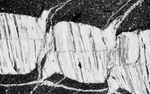
Figure 2. Fibrous calcite crystals with a high length to width ratio in a vein from a locality near Sestri Levante (Italy). Width of view is 10mm.
|
|
Figure 3. Fibrous quartz crystals between pieces of iron oxide from the Hamersley Ranges (Australia). Width of view is 8mm.
|

In this paper we demonstrate how the numerical models "Vein Growth" and "Fringe Growth" (Koehn et al., 2000; Bons, 2001) can produce different crystal textures found in natural veins and strain fringes and discuss implications for structural analysis. This paper is related to a number of papers that we published in recent years (Koehn and Passchier, 2000; Koehn et al., 2000, 2001; Bons, 2000, 2001 and Hilgers et al., 2001). A more general introduction and review than the one presented here is given in Bons (2000). The programs that we used for the simulations are described in detail in Koehn et al. (2000, "Fringe Growth") and Bons (2001, "Vein Growth"), a short introduction is given in chapter 2 of this paper. In Koehn et al. (2001) and Hilgers et al. (2001) we simulated the development of crystal textures in specific examples of natural fibrous veins and strain fringes. A more general approach is presented in chapter 3 including a simulation of a striped bedding-vein (Koehn and Passchier, 2000). The structural interpretation shown in chapter 4 is a visualization of the "object-centre path" method presented in Koehn et al. (2001).