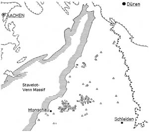
Data collected in this study come from about 150 outcrops containing mullion structures in lower Devonian shallow marine sands and shales (Fig. 5). In addition we report data from a smaller number of outcrops in the Bastogne area (Spaeth, 1986; Sippli, 1981).
Figure 5. Location of outcrops
Map showing the location of most of the outcrops used for the statistical analysis of this study. For location refer to Figure 2.
Most of our observations are consistent with earlier descriptions from the literature. We supplement these by a number of observations not yet reported. The observations are illustrated in a series of figures. Extensive descriptions are for example (Bruhl, 1969; Pilger and Schmidt, 1957a; Jongmans and Cosgrove, 1994; Sintubin et al., 2000; Corin, 1933; Mukhopadhyay, 1972; Rondeel and Voermans, 1975; Tromme, 1997)
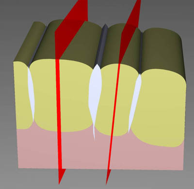
• Mullions are visible as highly cylindrical cuspate-lobate structures of exposed psammite-pelite interfaces (Fig. 6). The cusps always point towards the psammitic layer. The higher the lithological contrast, the better defined the mullion.
Figure 6. Characteristic structures of the Eifel-Ardennes mullions
(a) 3D model showing the major characteristic structures of the Eifel-Ardennes mullions. The morphology of the structures is not strongly dependent on size: width of this model can be between 20 cm and 5 m. Click for Movie
• Mullions are always associated with quartz-rich veins in the psammite layers, terminating close to cusps. This association is very strong, over 99% of all observed cases contain this association. In psammitic layers the quartz veins are better developed. Sometimes these intra-mullion veins cross the psammite-pelite interface and terminate inside the pelite layer.
Figure 7. Small outcrop near the village of Rouette, Belgium
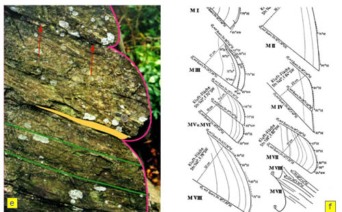
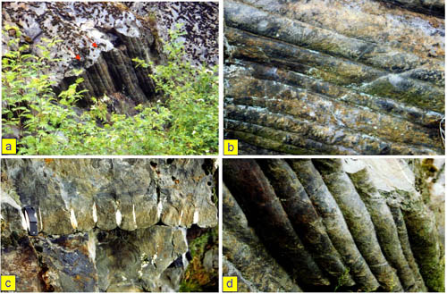
Features in a small outcrop containing dm-size mullions, near the village of Rouette, Belgium. In this outcrop the angle between mullion axis and delta-lineation is approximately 20 degrees (only vaguely visible on the photographs). (a) Overview of the outcrop exposing the bottom of a psammite layer embedded in pelite. The mullions are developed at two wavelengths, at the m and dm scale. (b) Bottom of a relatively clay-rich psammite layer, with mullions less well developed. Note the lateral termination of some of the cusps, transforming two mullions into one. Width of image is about 1 m. (c) Profile view of the main psammite layer, with well developed mullions. Every cusp in this outcrop has an associated quartz vein. Width of image is approximately 50 cm. (d) Bottom of main mullion-containing layer. The image contains one of the first order mullions, with 7 smaller ones superimposed. (e) Sample cut perpendicular to the mullion axis, showing details of cleavage development in the cusps, and deformation of the veins. Width of image is 20 cm.
In layers without veins no mullions are observed, but in the same outcrop veins can be present without mullions. (Fig. 7).
Mullions are found on both limbs and in the hinge zones of regional folds. In mullions, cleavage and bedding intersect at angles more than 40 degrees. Mullions do not occur on fold limbs where the cleavage is sub-parallel to bedding. In these uncommon cases the psammite layers show the "normal, extensional" boudinage with cleavage that follows the curvature of bedding in the boudin necks.
When the psammite layer's boundary is well defined and bounded by pelite on both top and bottom, mullions are formed with cuspate-lobate folds on both upper and lower interface. The lobes point away from the psammite layer, with the intra-mullion veins connecting the cusps. In layers with graded bedding mullions are present at one side of the layer only, with cusps associated with veins (Fig. 8).
Figure 8. Large outcrop near Bastogne
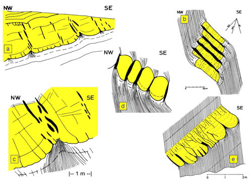
(a) Large outcrop along the railway line North of Bastogne, showing mullions developed at two wavelengths. Note that in the cusps between the large mullions the quartz veins are thicker, too. Width of outcrop 50 m. Slightly modified after Brühl (1969). (b) A series of small mullions showing the very slender aspect ratios sometimes observed. Slightly modified after Brühl (1969). (c and d) Mullions showing the fans of cleavage convergent into the cusps. Slightly modified after Brühl (1969). (e) Sandstone layer with graded bedding, showing mullions developed only at the contact with high material contrast. Slightly modified after Brühl (1969). (Click for enlargement)
Cleavage is well developed in the slate layers, and is refracted across layer boundaries. Near the cusps between mullions, cleavage forms well-developed fans convergent into the cusps (Fig. 8c) (Mukhopadhyay, 1972).
Composite mullions with two wavelengths are sometimes found, usually the cusps between the larger first order lobes are connected to thicker quartz veins (Fig. 8a, Fig. 7a).
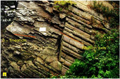
Mullion axes are oriented close to but usually not parallel to the delta lineation (Fig. 4e). Well-defined mullions with their axis at more than 40 degrees to delta lineation have not been documented. Delta lineation is rarely exactly parallel to mullion axis. Brühl (1969) quotes, based on 50 data, the angle between mullion axis and delta lineation to have values usually between 0 and 20 degrees. To further illustrate this point, outcrop data were grouped into subsets with similar structures. Figure 14 shows stereoplots of bedding, cleavage, mullion axis and delta lineation for two of these groups. It can be seen that in some outcrop groups the delta lineation is sub-parallel to the mullion axis, and in others the orientations are clearly different. In a compilation plot (Fig. 15) of all mullion axes and delta lineations, this distinction is not so clear due to the regional trends in orientations. For all outcrops we calculated the angle alpha between delta lineation and mullion axis. Figure 16 is a histogram of these data. There is a clear maximum at around 20 degrees, with no reliable measurement over 30 degrees. Frequency also decreases towards low values of alpha, but less rapidly than for high angles. This is consistent with the observed correlation between azimuth of mullion axis and delta lineation (Fig. 16). In rare cases mullions are folded across small-scale folds with the fold axis at an angle to the (folded) mullion axis (Bruhl, 1969), consistent with the above observation.
Figure 9. Microstructure of thin quartz vein
a) Microstructure of thin quartz vein at high angle to bedding, in a thin psammite layer containing mullions. b) Enlargement of this microstructure shows stretched crystals. (Click for enlargement)
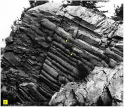
In cross section, mullions show layering which is straight in the middle of the psammite layer, with the curvature increasing towards the outside. Bedding planes in the adjacent pelite layer follow the cuspate-lobate shape until they are about one mullion-amplitude away from the layer contact (Fig. 7e). In thin sections of the psammite layers in mullions, the amount of solution-precipitation deformation is highest in the centre of the layer (Fig. 10) (Spaeth, 1986).
Figure 10. Microstructure in a small mullion

Microstructure of psammite at different locations in a small mullion. The amount of inferred solution transfer in the middle of the layer is higher than in the layer's boundary. (Click for enlargement)
Layer thickness correlates with the width of mullions (Jongmans and Cosgrove, 1994; Rondeel and Voermans, 1975). The aspect ratio H/W usually between 1 and 3 (Fig. 13). Several authors have noted that this is too slender in comparison with “normal” boudins formed by layer-parallel extension.
Mullions have orthorhombic symmetry when cleavage is normal to bedding, and are monoclinic on fold limbs (Fig. 4). In this case, symmetry is in agreement with the corresponding fold limb.
Mullions are highly cylindrical, with scatter in mullion axes typically less than 15 degrees in one large outcrop. Observations on cylindricity over more than 10 metres are much less frequent, as such outcrops are rare. In more detail, cusps between mullions sometimes end along strike, so that two mullions join into one (Fig. 7d, Fig. 13e).
Figure 11. Mullions with veins
Mullions with veins, which are clearly stretched at high angles to bedding, with the development of boudins. Note the difference in aspect ratio between the boudins and mullions. Slightly modified after Brühl (1969). (Click for enlargement)
Measured along the layering, veins comprise up to about 10% of bed length. This ratio gradually decreases towards the vein’s tip.
Veins between mullions are normally spindle- or lens shaped. They are generally oriented at high angle to bedding and in profile view are sub-parallel to the weakly developed fracture cleavage in the psammite layers.
Vein microstructure is variable: in some cases fibres parallel to bedding are found. In other cases, the quartz has a blocky microstructure (Fig. 10).
Figure 12. Microstructure of a deformed intra-mullion quartz vein showing evidence for crystal plasticity and incipient recrystallization. (Click for enlargement)
In thin sections, micro-scale veins parallel to the intramullion veins are occasionally found, with a stretched crystal microstructure indicating transgranular fracturing in a strong rock (Hilgers et al., 2000) (Fig. 9, Fig. 11).
Not unfrequently the intra-mullion veins have a pinch-and-swell structure (stretched in a direction sub-normal to bedding); these structures have the usual aspect ratios and are quite different from the Mullions (Bruhl, 1969; Jongmans and Cosgrove, 1994).
In thin sections of deformed veins the vein quartz shows evidence for plastic deformation, with subgrain formation and incipient recrystallisation (Fig. 12).
Figure 13. Structures from the Bastogne quarry
Structures from the Bastogne quarry. (a) Mullions developed in a thick psammite layer. Note the unusual aspect ratios. Width of outcrop about 10 m. (b) Detail of (a). (c) Regular series of mullions in a thick sand layer. Width of outcrop is approximately 10 m. (d) Detail from a mullion similar to (c), showing the convergent cleavage fan in the cusp. Width approximately 10 cm. (e) Photograph of the bottom of a psammite layer, showing cylindrical mullions. Note cleavage approximately parallel to bedding. Note the lateral termination of the mullions. Width of outcrop about 15 m.
Fluids in intramullion veins have variable composition but can be H2O, N2 and/or CO2 – rich as has been documented in extensive studies (Fielitz and Mansy, 1999; Sintubin et al., 2000; Schroyen and Muchez, 2000; Darimont et al., 1988).